Cyanotic congenital heart disease with increased pulmonary blood flow
(1-2) L-transposition of the great arteries (Congenitally corrected TGA, L-TGA)
Classified as {S, L, L} in segmental sequential analysis and there is no cyanosis in this disease on birth (due to its hemodynamically normal condition without cardiac anomaly).
Blood flow in congenitally corrected TGA
– Systemic circulation ➔ Right atrium ➔ Left ventricle ➔ Pulmonary circulation ➔ Left atrium ➔ Right ventricle ➔ Systemic circulation …
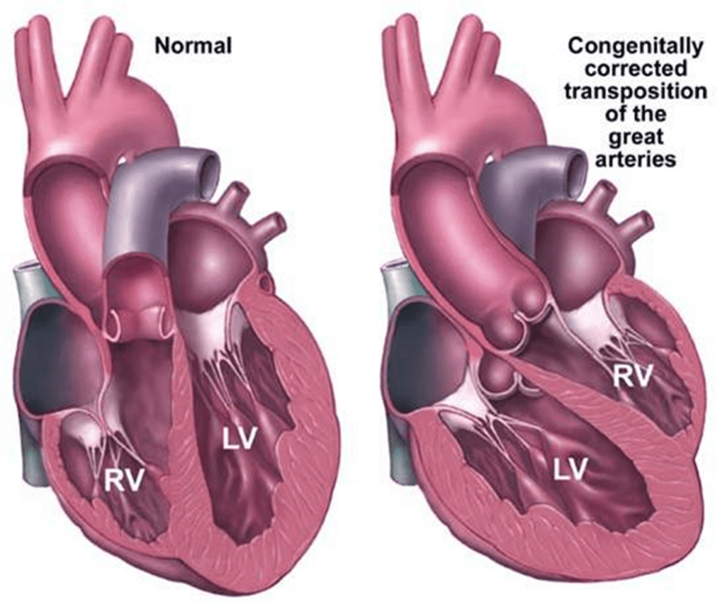
The definition of congenitally corrected TGA
Congenitallcy corrected TGA (also called as L-transposition of the great arteries, L-TGA) is a spectrum of cardiac malformations where the atrial chambers are joined to morphologically inappropriate ventricles, and the ventricles then give rise to morphologically inappropriate arterial trunks.
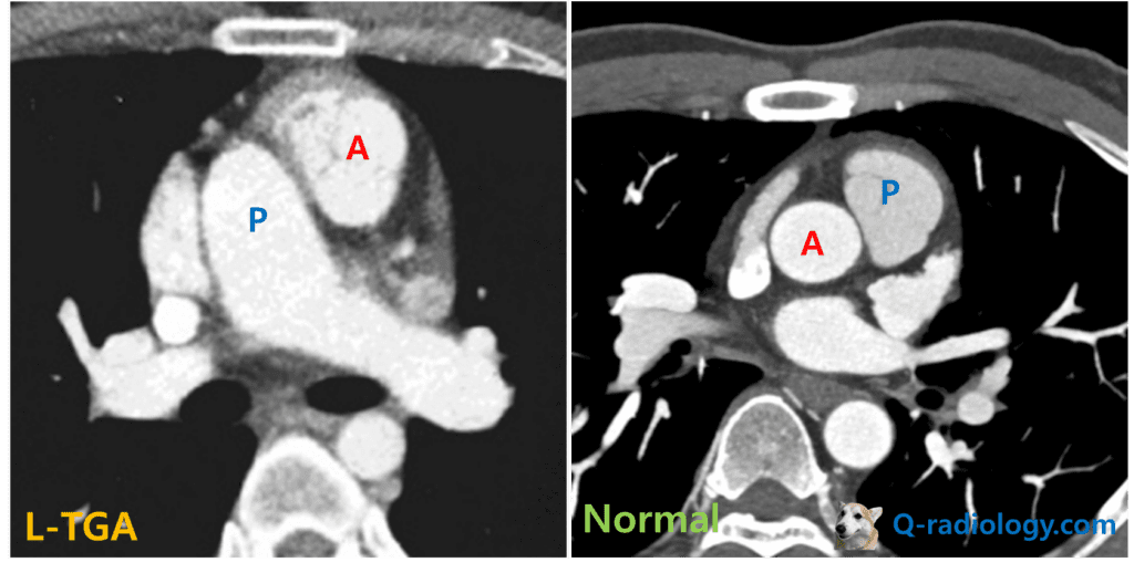
In ccTGA (as well as D-TGA), the pulmonary artery and the aorta take a parallel course, in contrast to the crossing configuration seen in the normal heart.
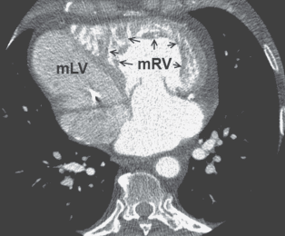
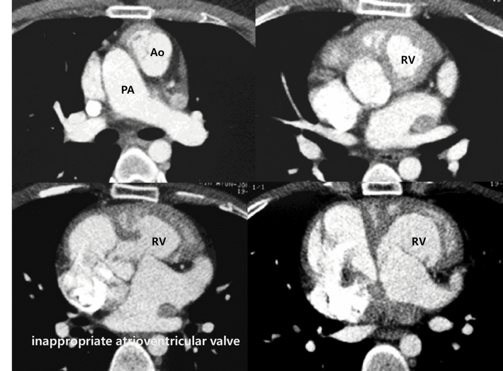
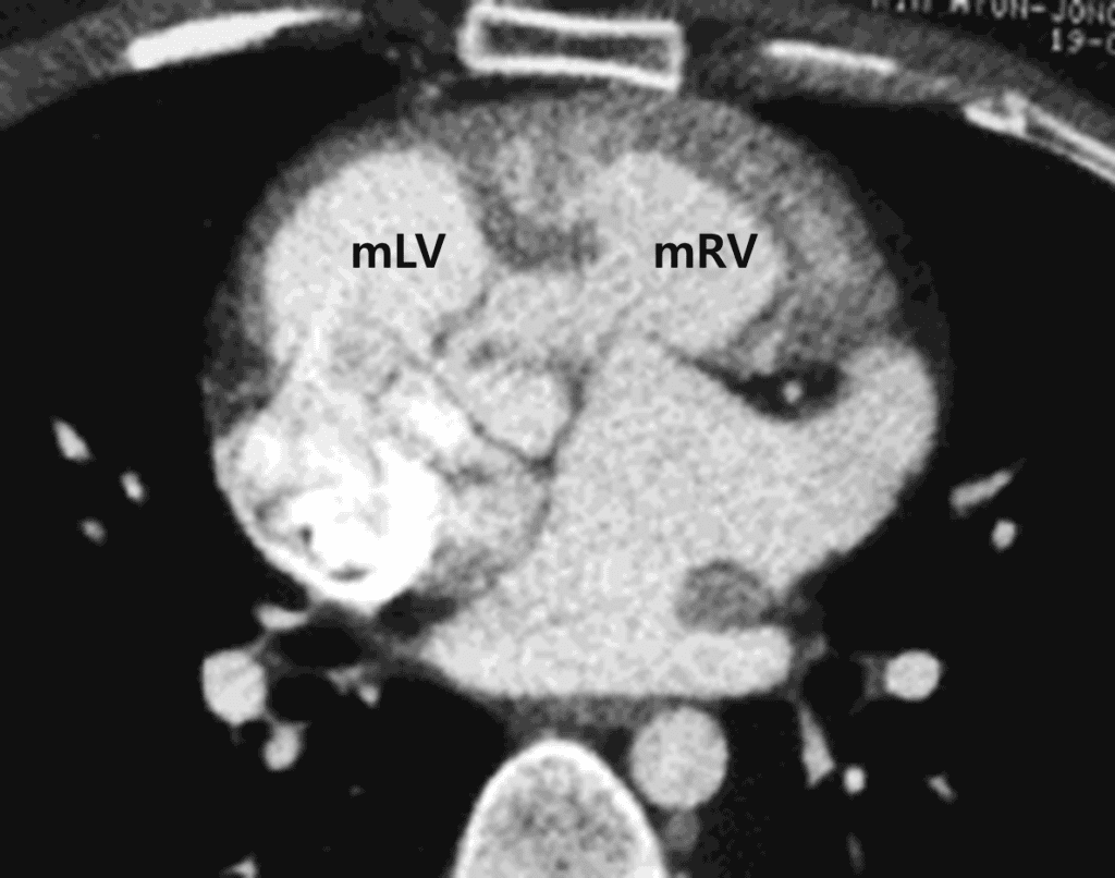
Find morphological RV in imaging to diagnose ccTGA
The RV can be recognized by its coarse trabeculated myocardium, the moderator band, the presence of septal attachments for the papillary muscles, and more apically displaced insertion of the AV valve on the interventricular septum.
Treatment of ccTGA
(1) Pulmonary artery banding : to reduce the pulmonary blood flow
(2) LV to PA conduit : when pulmonary stenosis exists
(3) Double switch operation : atrial switch and arterial switch
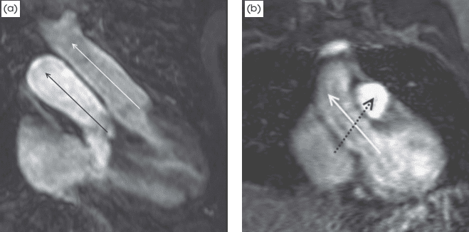
Note the parallel course of the pulmonary artery (black arrow) and the aorta (white arrow). Contrast this with (b), which is a coronal MRA image of a normal heart. Note the crossing pattern of the aorta (white arrow) and the pulmonary artery (dotted black arrow), which crosses posterior to the plane of this image.
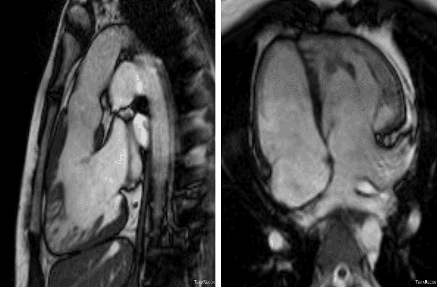
Right) Morphologic RV is located in left side
Reference)
Charles S. White, Linda B. Haramati, Joseph Jen-Sho Chen, and Jeffrey M. Levsky (2014), Cardiac Imaging, Oxford university press
Related posts:
 Tetralogy of Fallot : Cyanotic congenital heart disease with decreased pulmonary blood flow
 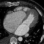 Ebstein anomaly : Cyanotic congenital heart disease with decreased pulmonary blood flow
  Tricuspid atresia : Cyanotic congenital heart disease with decreased pulmonary blood flowTricuspid atresia
  Transposition of the great arteries (D-TGA)