Castleman disease radiology

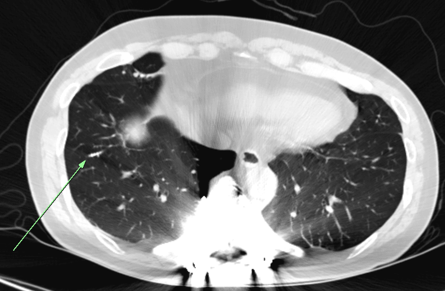
DEFINITIONCastleman disease is an uncommon benign B-cell lymphoproliferative disorder. TERMINOLOGY= angiofollicular lymph node hyperplasia = giant lymph node hyperplasia 1) Hyaline vascular type (90%) – Unicentric castlman disease – Before 30s, no symptom – Single mass or mediastinal/hilar lymph node enlargement – Localized disease – No progression to non-hodgkin lymphoma – Mediastinal or hilar mass/LNE … Read more

Pulmonary hamartoma radiology

DEFINITION – Benign pulmonary tumor containing multiple mesenchymal tissue elements PATHOLOGY – Most common benign lung tumor (75%) – Common in 40-50s – Varying amounts of cartilage, fat, connective tissue, smooth muscle … IMAGING – Peripheral location (2/3) – Endobronchial location (10%) ; obstructive symptom – Round or smooth lobulated mass with popcorn calcification and … Read more

Paradoxical response in tuberculosis : radiology

DEFINITION New lung lesions that develop during medication for tuberculosis which should be considered a transient worsening that ultimately improves with continuation of medication IMAGING – This condition occurs in 2 to 12 weeks of treatment – Peripheral lung nodule or mass (1~8cm) ipsilateral to pleural effusion – Heterogenous enhancing nodule/mass, no air-bronchogram or no … Read more

Pneumocystis jirovecii pneumonia radiology

DEFINITION Opportunistic fungal infection that affects individuals with T-cell immunodeficiency IMAGING – Radiograph ○ Normal, diffuse bilateral heterogeneous opacities ○ Spontaneous pneumothorax in HIV(+) patient is nearly diagnostic of PCP – CT ○ Ground-glass opacities are dominant finding ➜ Most commonly central with relative subpleural sparing (41%) ➜ Heterogeneous with mosaic attenuation (29%), or diffuse … Read more

Chronic eosinophilic pneumonia radiology

DEFINITION Idiopathic lung disease characterized by respiratory symptoms > 2 weeks, parenchymal opacities, and marked tissue and peripheral blood eosinophilia CLINICAL FINDING – Mid age female (>40) – 2/3 of patients have prior history of asthma – 1/2 of patients have history of atopy – Not related with smoking – Insidious clinical course with average … Read more

Superior Vena Cava syndrome radiology

DEFINITION – Superior vena cava syndrome (also called SVC obstruction) is a group of symptoms that occurs when SVC is obstructed by intraluminal or extrinsic disease. – SVC syndrome is characterized by Impaired venous return from head, neck, upper extremities, and trunk CLINICAL FINDING– Face, neck, upper trunk, and upper extremity edema IMAGING CT and … Read more

Relapsing polychondiritis radiology : smooth anterolateral tracheal wall thickening

DEFINITION Autoimmune disease characterized by recurrent inflammation of cartilaginous structures CLINICAL FINDING – Common in 20~30s, but all age group are possible – Auricular chondritis – Ocular manifestations (scleritis, episcleritis, or conjunctivitis) – Nasal chondritis (painful inflammation of nasal cartilage) – Costochondritis (retrosternal chest pain) – Laryngotracheal and pulmonary involvement (50% during disease course) – … Read more

Tracheobronchopathia osteochondroplastica (TBO) radiology : nodular tracheal wall thickening

DEFINITIONTracheobronchopathis osteochondroplastica (TBO) is a rare disease with unknown etiology characterized multiple submucosal osteocartilaginous nodules in central airway walls (trachea and proximal bronchi).multiple nodules protrude into lumen of the airways with narrowing it CLINICAL FINDING – Male, 50s – Dyspnea, hoarseness, cough, hemoptysis, recurrent lung infection IMAGINGCT – Multiple mural nodules with or without calcification … Read more