1. Atrial situs : SIA
Situs Solitus (Normal)
Situs Inversus (usually with Mirror Image Dextrocardia)
– Left-right reversal of the cardiac chambers coupled with a right sided stomach, right apex, and usually right arch.
– A slightly increased risk (3-5%) of congenital heart disease (Most commonly corrected transposition)
– Patients with Kartagener’s syndrome (Situs inversus with mirror image dextrocardia)
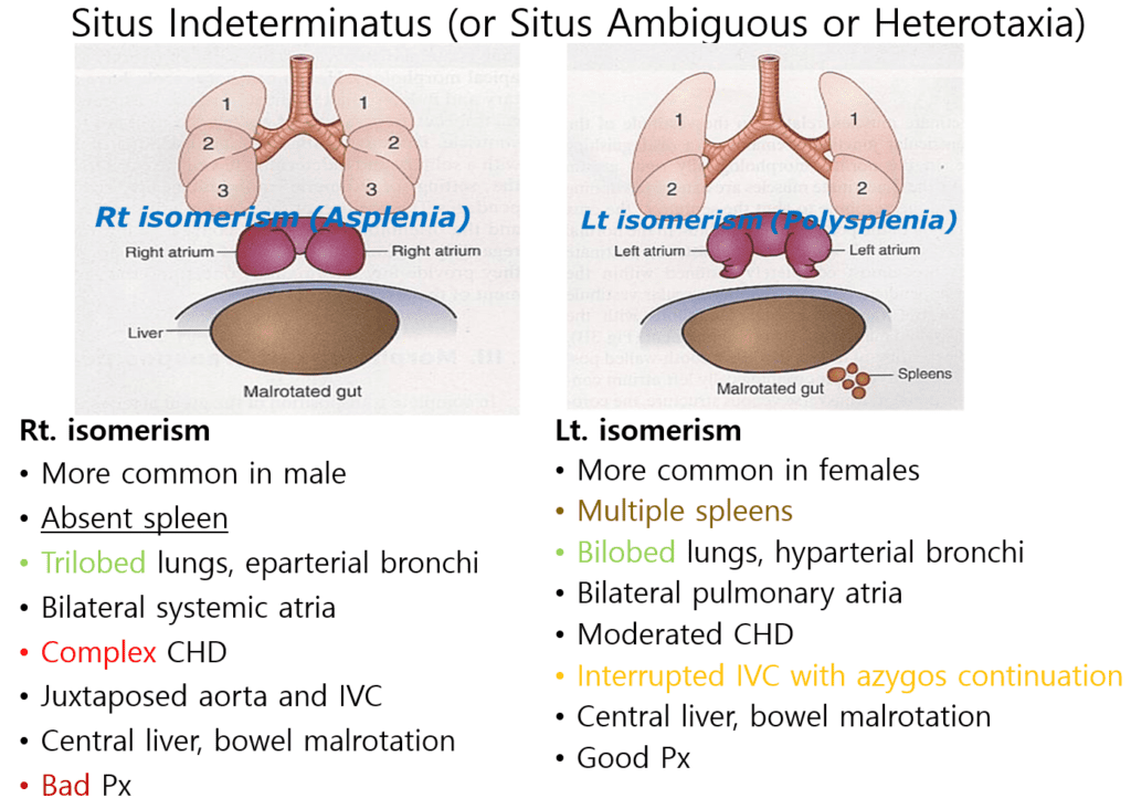
Situs Indeterminatus (or situs ambiguous or heterotaxia)
– Abnormal arrangement of the organs and vessels.
– Stomach and liver are typically midline
– Asplenia or polysplenia
– Congenital heart disease occurs in 50-100% of cases.

2. Ventricular loop (D/L-loop)
Septomarginal trabeculation – morphologic right ventricle
IF the septomarginal trabeculation is located in right side ? = D-loop
IF the septomarginal trabeculation is located in left side ? = L-loop
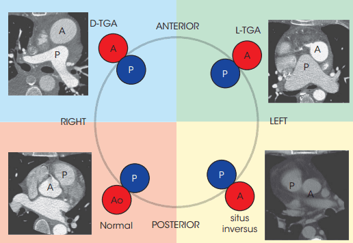
3. Great artery relationship (SIDL)
Solitus, Inversus, D-transposition, L-transposition
Reference)
Charles S. White, Linda B. Haramati, Joseph Jen-Sho Chen, and Jeffrey M. Levsky (2014), Cardiac Imaging, Oxford university press