Today’s case 11 – May-Thurner syndrome
Today I will introduce May-Thurner syndrome induced DVT and its treatment
I’m not sure why, but lately there seems to be an unusually high number of patients with Deep Vein Thrombosis (DVT) caused by May-Thurner Syndrome.
Personally, I think it might be related to the current cold weather in Korea. People are likely spending more time indoors and engaging in less physical activity, which could possibly contribute to a higher incidence of DVT.
This is just a speculation, though, without any solid evidence.
The patient is 53-year-old male.
The patient was referred to our facility following a diagnosis of Deep Vein Thrombosis (DVT).
Their primary concern was the sudden onset of swelling in the left lower limb, which had been present for two days.
The patient reffered for interventional procedure to interventional radiology department.
Consistent with our standard protocol, plans were made for the insertion of an IVC filter, thrombectomy, and stenting of the left common iliac vein.
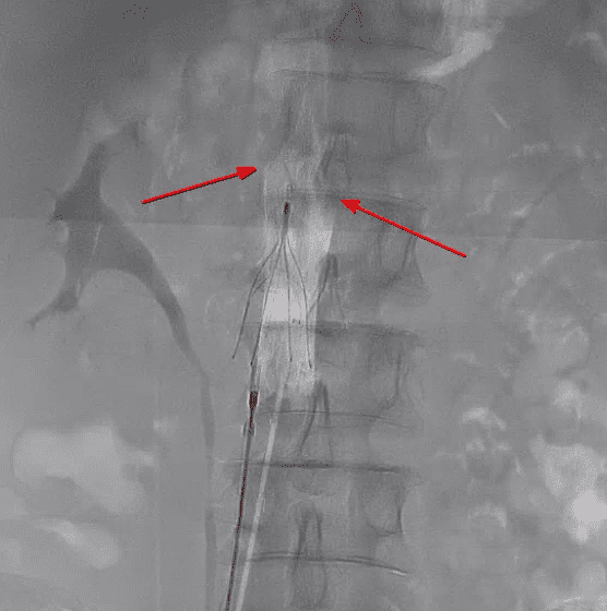
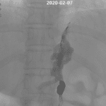
After puncturing the left popliteal vein, a 5F multipurpose catheter was introduced through a 9F sheath, chosen to accommodate subsequent stent insertion.
Upon gentle injection of contrast, an extensive filling defect indicative of thrombosis was observed.
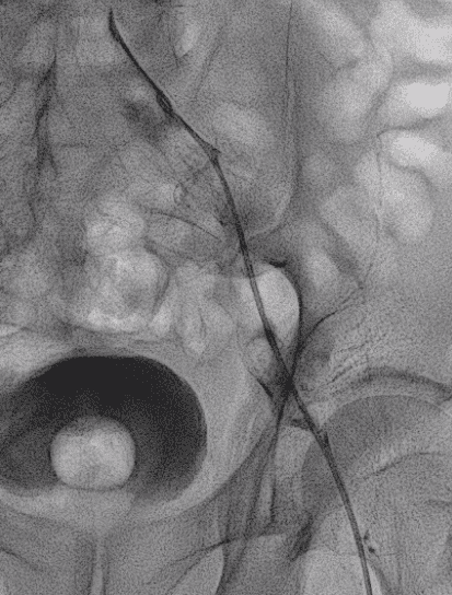
We utilized the AngioJet™ Peripheral Thrombectomy System from Boston Scientific for the procedure.
A power pulse injection of recombinant tissue plasminogen activator (r-tPA) was administered, followed by a 10-minute waiting period to allow for effective thrombolysis, which is essential for the subsequent aspiration of the thrombus.
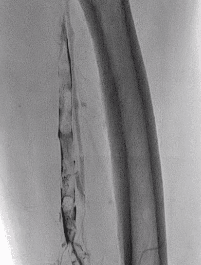
Finally, rheolytic thrombectomy was carried out from the proximal to the distal sections, starting from the left common iliac vein and extending to the left popliteal vein


This is likely due to chronic thrombus content, rather than other thrombi located below the left common iliac vein.


Plus
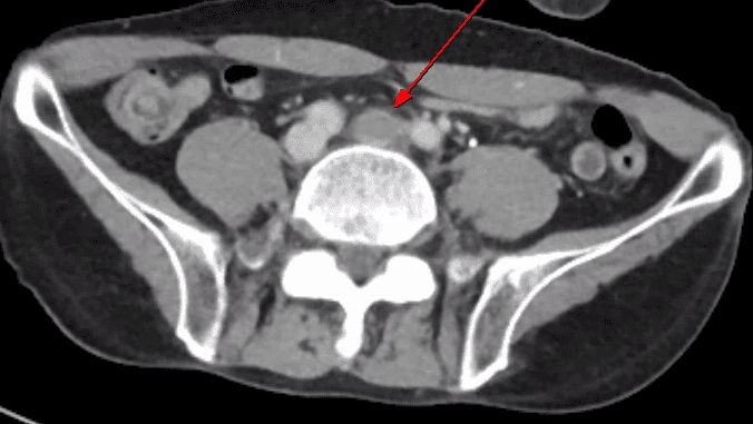
What is the May-Thurner syndrome ?

Keep in mind that this condition involves the collapse of the left common iliac vein due to external compression by the right common iliac artery and the vertebra
Instagram : Visit my instagram
Q-radiology : Home
Hloow.com : My korean website
Related posts:
 Coil Assisted Retrograde Transvenous Obliteration (CARTO): Treatment of variceal bleeding with interventional procedure (CARTO)
 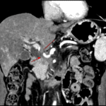 Today’s case 2 – Portal vein stent insertion for invaded main portal vein by pancreas head cancer
  Today’s case 3 – PICC (peripherally inserted central catheter)
  Today’s case 4 – Iatrogenic bleeding of deep femoral artery while insertion of A-line