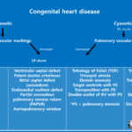
Cyanotic congenital heart disease with increased pulmonary blood flow
(1-1) D-transposition of the great arteries (Complete TGA)
D-transposition of the great arteries
D-transposition of the great arteries (D-TGA), also called complete transposition, causes cyanosis in infancy, which results from the aorta arising from the morphological right ventricle and the pulmonary artery arising from the morphological left ventricle.
The atrioventricular connections remain normal.
Since the systemic and pulmonary circuits are in parallel arrangement, a shunt such as a patent ductus arteriosus (PDA), ventricular septal defect (VSD), which is most common, or atrial septal defect (ASD) must be present for survival.
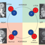
In D-TGA, the aortic root is anterior and to the right of pulmonary artery root, in contrast to the normal situation, where the aorta is posterior and to the right.


Systemic circuit In D-TGA(deoxygenated blood)
IVC, SVC ➔ Right atrium ➔ Right ventricle ➔ Aorta ➔ Whole body ➔ IVC, SVC
Pulmonary circuit In D-TGA(oxygenated blood)
Pulmonary vein ➔ Left atrium ➔ Left ventricle ➔ Pulmonary artery ➔ Lung ➔ Pulmonary vein
This parallel arrangement of the systemic and pulmonary vascular systems can only sustain life if there is enough mixing of blood via shunts (ASD, VSD, and PDA).
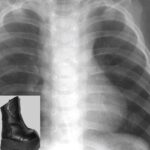
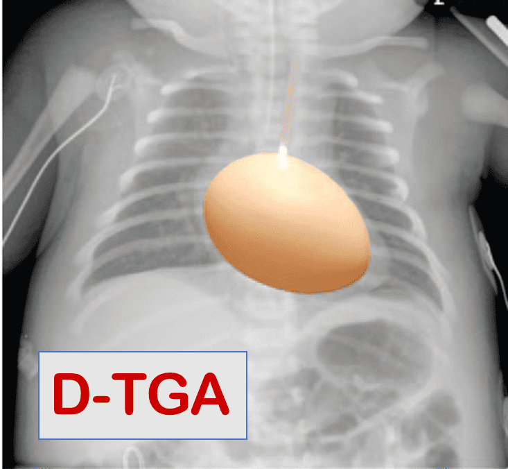
D-TGA on chest radiograph
Egg-on-string sign
– AP relation of great arteries
– Narrow superior mediastinum
– Round right border of the heart
Treatment of complete TGA
1) Jatene procedure (Arterial switch)
– The current treatment of choice
2) Mustard or Senning procedure (Atrial switch)
– Previous surgeries of choice for D-TGA were Mustard and Senning procedures, which restored a serial circulation at the atrial level.
– Complications : 1) superior baffle stenosis 2) baffle leak 3) RV dilatation and dysfunction
3) Rastelli procedure
– This procedure is possible when there is D-TGA and a well-positioned VSD
– Oxygenated blood from the left ventricle is routed (“baffled”) through the VSD into the aorta, such that the left ventricle is the systemic pump. The pulmonary artery is ligated proximally and an extracardiac conduit is then constructed directly from the right ventricle to the pulmonary artery. The conduit transmits deoxygenated blood from the right ventricle to the pulmonary arteries

Image from Angeline D. Opina and Wayne J. Franklin. D-Transposition of the Great Arteries: A New Era in Cardiology. CVIA. Vol. 3(1):85-92. DOI: 10.15212/CVIA.2017.0037


a) Preoperative view of heart with transposition, VSD, and LVOT obstruction
b) Intraoperatively, left ventricular blood flow has been redirected through the VSD to the aorta, and main PA has been transected
c) Extracardiac valved conduit from right ventricle to Pas has been creasted
Reference)
Charles S. White, Linda B. Haramati, Joseph Jen-Sho Chen, and Jeffrey M. Levsky (2014), Cardiac Imaging, Oxford university press