F/71
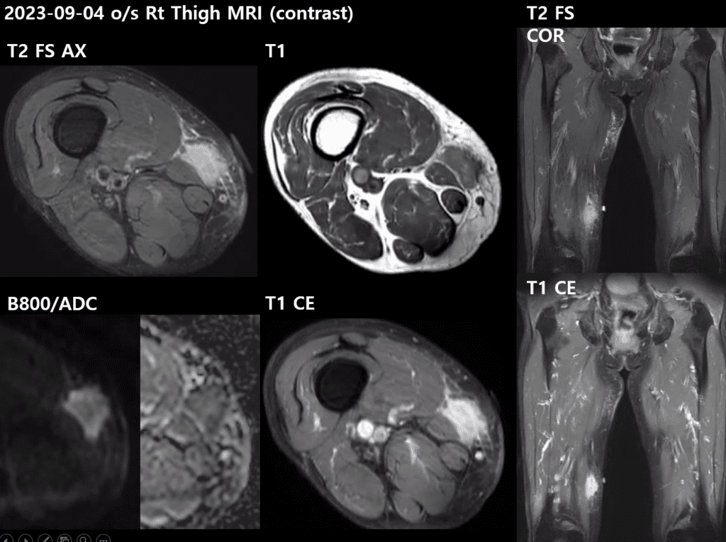
C.C.> Rt thigh mass (2MA)
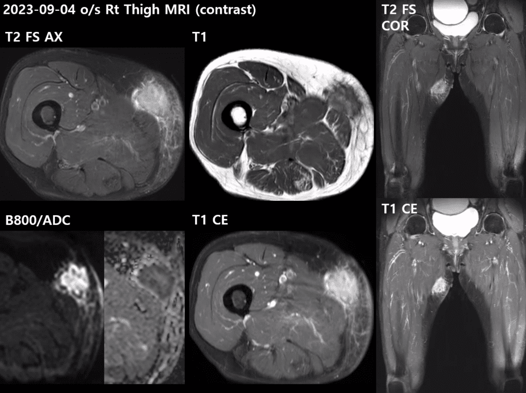
– 3.5 x 1.9 x 3.3 cm
– T2 high, T1 heterogeneous high~isointense SI
– closely abutting the nearby deep superficial fascia, but no direct intramuscular tumor invasion into the adductor longus and gracilis muscles.
– localized thickening of the overlying skin
– very dirty fat stranding (+)
– showing heterogeneous avid contrast enhancement
– DWI/ADC: marked diffusion restriction (+)

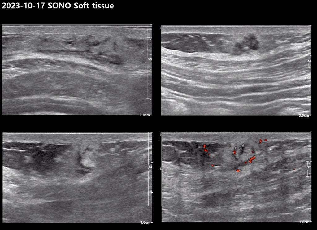
– increased vascularity
– not compressible
– no tenderness
– similar morphology with thigh mass
| Aspect | Details |
|---|---|
| General Description | Sarcoidosis: Systemic granulomatous inflammatory disorder |
| Muscle Lesions | Occur in 50-80% of sarcoidosis patients; symptomatic in only 0.5-2.5% |
| Types of Muscular Sarcoidosis | |
| Chronic Sarcoidosis Myopathy | Most common (86%), CT/MRI usually normal, occasional muscle atrophy with fatty degeneration |
| Acute Sarcoidosis Myositis | Symmetrical proximal myalgia, MRI may be normal, can progress to muscle contractures with hypertrophy |
| Nodular Sarcoid Myositis | Multiple muscle nodules, preferentially located in lower limbs |
| Imaging Features of Nodular Sarcoidal Myopathy | |
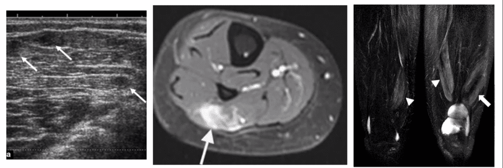
| USG (Ultrasound) | Nodules with hyperechoic center and hypoechoic periphery compared to adjacent muscles |
| MR (Magnetic Resonance) | Often at musculotendinous (MT) junction, multiple, bilateral, lower extremity (most common) |
| Dark Star Appearance (Axial): Bright rim with internal low signal intensity (SI) at T2WI and T1WI-CE | |
| Three Stripe Sign (Coronal/Sagittal): Inner decreased SI and outer stipple of high SI | |
| Pathology Correlation | Central area: Sarcoid nodule, dense fibrotic tissue; Outer area: Active inflammatory granuloma with epithelioid cells |