Heterotopic gray matter radiology

** Neuronal migration disorder ** Nodular heterotopias – subependymal heterotopia: most common– subcortical heterotopia Diffuse heterotopias – band heterotopia: Now this entity is classified as a spectrum of lissencephaly ; also known as double cortex heterotopia and X-linked lissencephaly (chromosome Xq22.3) ; double cortex sign : thin interface of WM between the band heterotopia and … Read more

METACHROMATIC LEUKODYSTROPHY Radiology

• Lysosomal storage disorder caused by ↓ arylsulfatase A (ARSA) resulting in central (CNS) and peripheral (PNS) nervous system demyelination• 3 clinical forms: Late infantile (most common), juvenile, adult IMAGING• Best diagnostic clue: Confluent butterfly-shaped ↑ T2 signal in deep cerebral hemispheric white matter◦ Early: Spares subcortical U-fibers◦ Late: Involves subcortical U-fibers• Sparing of perivenular … Read more

Cystic hygroma radiology

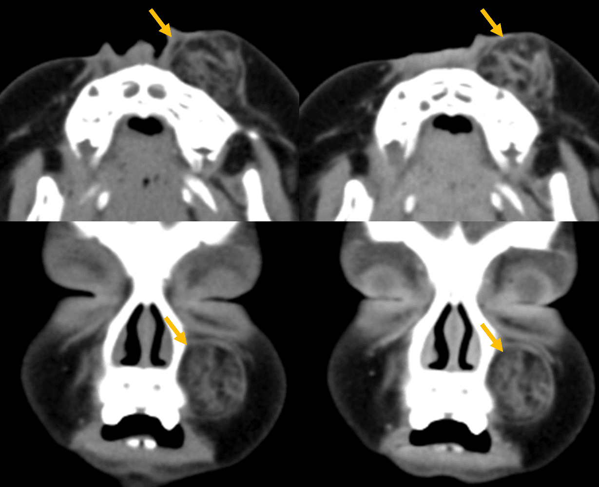
The term ‘cystic hygroma’ is corrected to macrocystic lymphatic malformation by ISSVA (International Society for the Study of Vascular Anomalies) IMAGING – Macrocystic lymphatic malformation ; Multiloculated cystic neck mass with imperceptible wall, thin septations, and fluid-fluid levels ; Location : Any face/neck location (not intracranial) ; Often in multiple contiguous spaces – transspatial ; … Read more

Epiglottitis radiology

Airway obstruction secondary to inflammation of epiglottis & surrounding tissues IMAGING – Lateral radiograph 1. Marked thickening of epiglottis 2. Thickening of aryepiglottic folds – Extend from epiglottis anteriosuperiorly to arytenoid cartilages posteroinferiorly – Normally thin and convex inferiorly – May become thickened and convex superiorly – Swelling of these folds ➜ Actual airway obstruction … Read more