Normal uterine anatomy on ultrasound/MRI
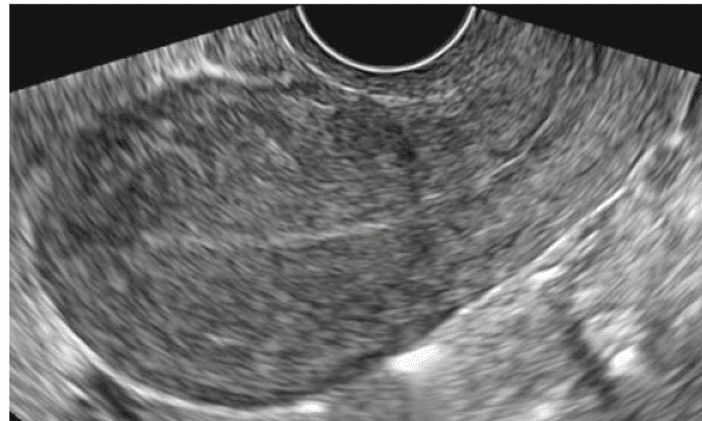
Uterine anatomy on MRI Uterine anatomy on TVUS (transvaginal ultrasound) See other posting about Genitourinary imaging Follow my instagram
Uterine anatomy on MRI Uterine anatomy on TVUS (transvaginal ultrasound) See other posting about Genitourinary imaging Follow my instagram
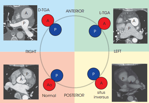
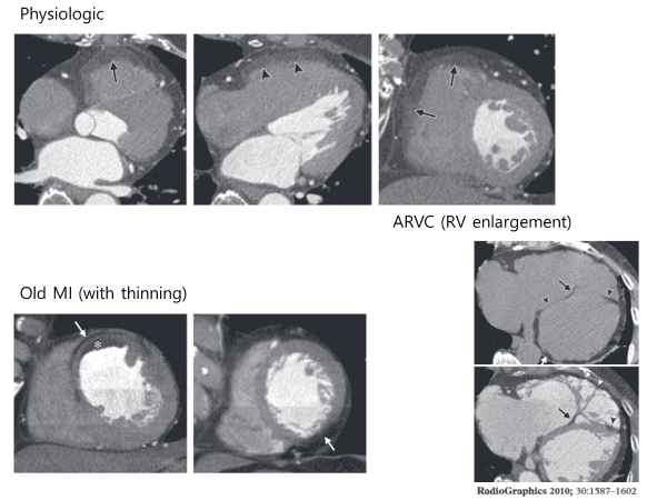
1. Atrial situs : SIASitus Solitus (Normal)Situs Inversus (usually with Mirror Image Dextrocardia) – Left-right reversal of the cardiac chambers coupled with a right sided stomach, right apex, and usually right arch. – A slightly increased risk (3-5%) of congenital heart disease (Most commonly corrected transposition) – Patients with Kartagener’s syndrome (Situs inversus with mirror … Read more
Follow my instagram See other posting about cardiovascular imaging

The great cardiac vein•The main tributary of the coronary sinus•Begins near the apex of the heart→ Ascends with the anterior interventircular branch of the LCA (along LAD)→ At the coronary groove turn left runs around the left side of the heart with the circumflex branch (along LCX)→ to reach the coronary sinus
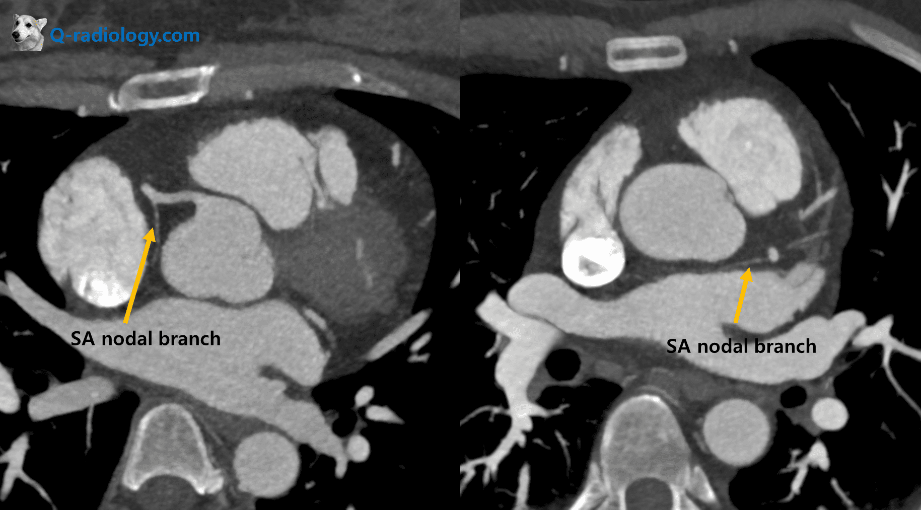
The sinoatrial nodal artery is the second branch that arises from the proximal RCA in 65.4%, immediately distal to the RCA origin. In 16.6% of cases, the sinoatrial nodal artery arises from the LCX
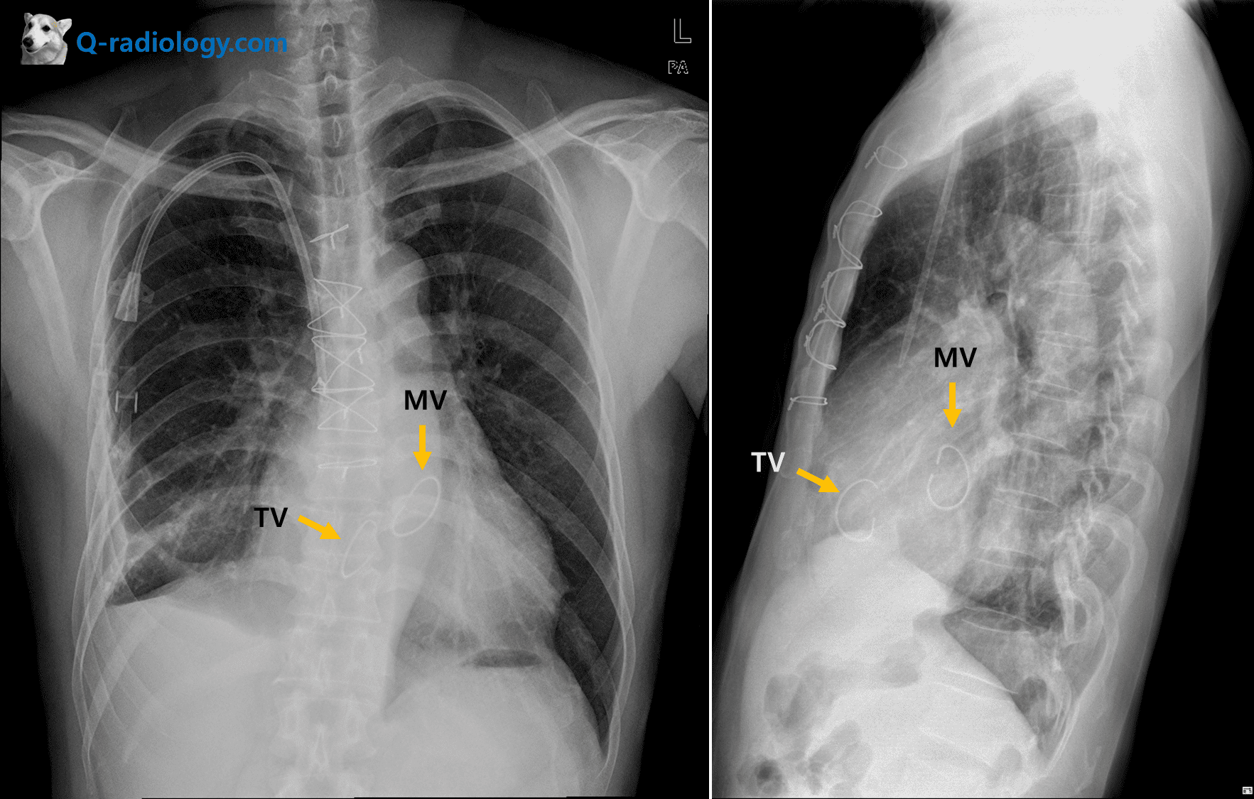
Valve anatomy on chest x-rayAortic valvePulmonary valveTricuspid valveMitral valve
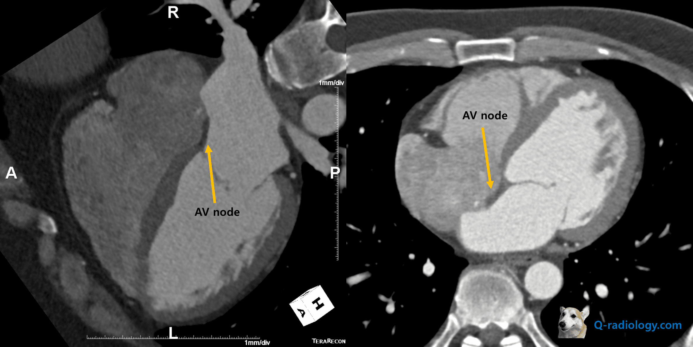
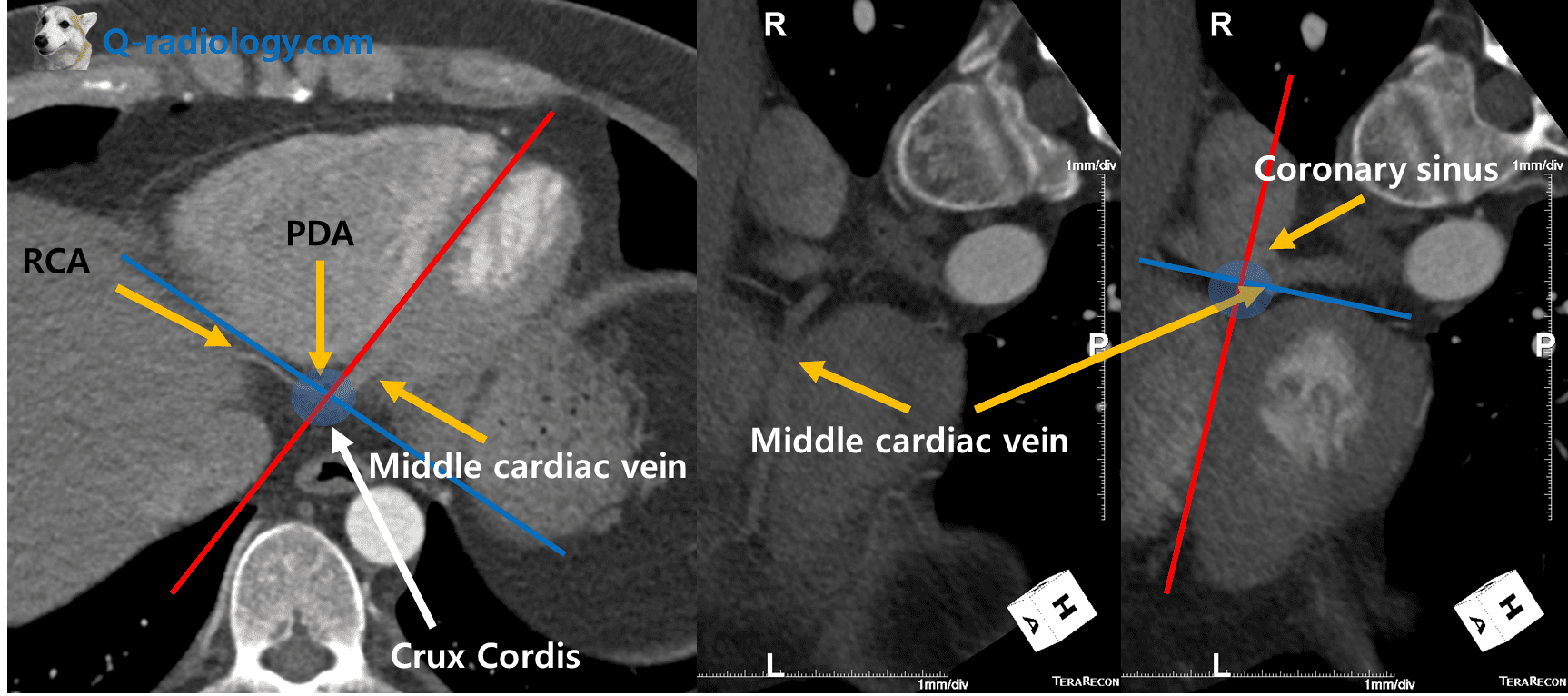
•Tendons of todaro•Coronary sinus•Tricuspid annulus Koch triangle contains AV node and His bundle See more about cardiovascular anatomy and radiology Follow my instagram
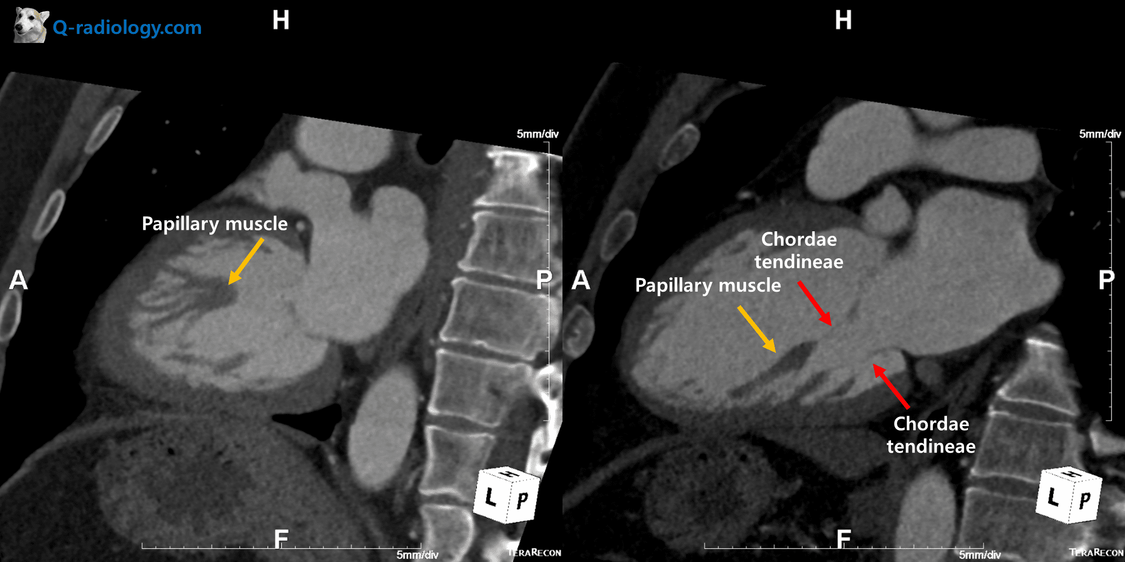
There are three papillary muscles in the RV, the anterior, posterior, and septal, and two papillary muscles in the LV, the anterolateral and posteromedial. Papillary muscles originate from the inner wall of the ventricles in their middle or apical portions, have an elongated and tapered trunk, and continue as chordae tendineae, which then attach to … Read more