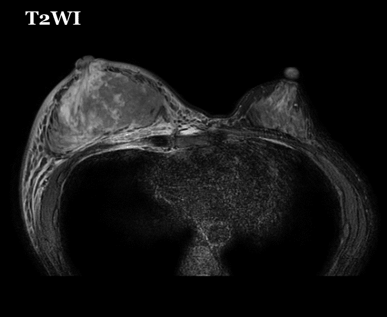
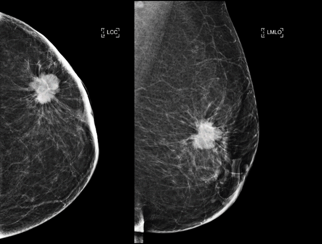
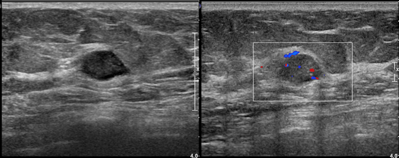
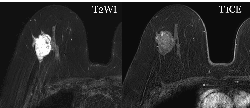
Breast cancer staging

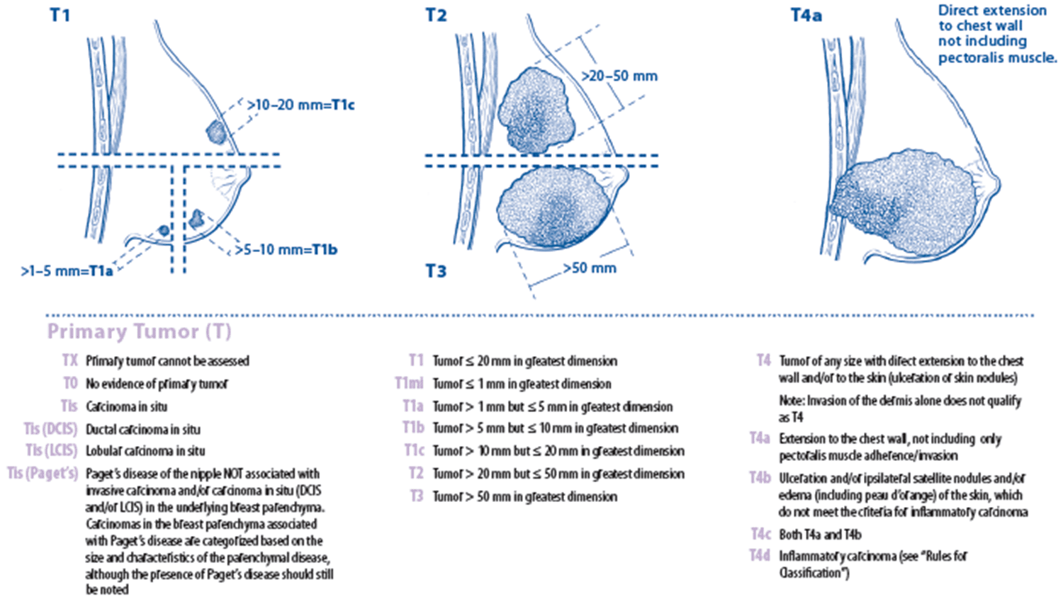
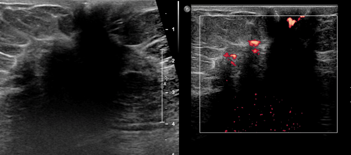
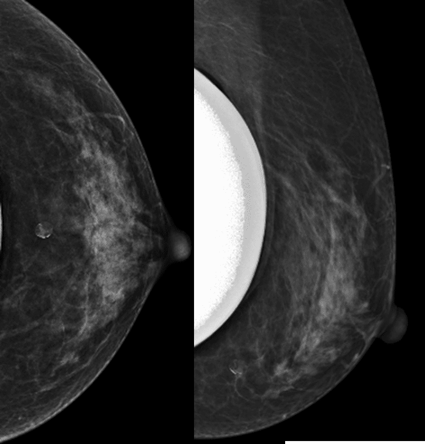
Determining Tumor Size Only the invasive component Identified specific imaging  modalities including MMG, US and MRI Tx T0 Tis Carcinoma in situ, with no evidence of an invasive component. Tis(DCIS) Tis(LCIS) Tis(Paget’s) : Paget’s dz of the nipple NOT associated with underlying breast cancer T1 Tumor ≤ 20mm in greatest dimension T1mi : microinvasion ≤ … Read more

Mucinous carcinoma of the breast: Breast cancer which shows high signal intensity on T2WI

Common in older age than IDC NOS Slow growing, more favorable prognosis DCIS are found in 75% of the patients Imaging finding US : Circumscribed, isoechoic or slightly hypoechoic mass, posterior enhancement > 50% MR : High signal intensity in T2WI due to mucinous component Let’s see a case (83 year-old female) from my hospital … Read more

Medullary carcinoma of the breast: Breast cancer which mimics the benign one

Younger women than IDC NOS More favorable prognosis Could be misdiagnosed as ‘fibroadenoma’ Calcification – Rare Imaging finding MG : partially circumscribed mass, calcification rare US : hypoechoic mass with posterior enhancement Let’s see a case (33 year-old woman) from my hospital confirmed as medullary carcinoma in CNB Other Circumscribed masses Medullary carcinoma, Papillary carcinoma, … Read more

Pregnancy-associated breast disease

1) mastitis/abscess 2) Galactocele 3) Lactating adnoma ➤ Most breast masses that develop during pregnancy and lactation are benign 4) Pregnancy associated cancer Breast malignancy diagnosed during pregnancy or after pregnancy upto one year Accounts 0.2 to 3% of all breast cancers Young age, associated with BRCA gene mutation Most PABC are in advanced stage … Read more

Fat containing masses on mammography: Malignant lesion or benign one?

1) Fat necrosis Lucent mass with dystrophic calcifications Associated with surgery or trauma Follow up in 3-6 months : typically decrease in size 2) Oil cyst An area of focal fat necrosis becomes walled off by fibrous tissue. 3) Galactocele : Fat fluid level on 90′ lateral view M/C benign breast lesions in lactating women … Read more