Splenic infection radiology

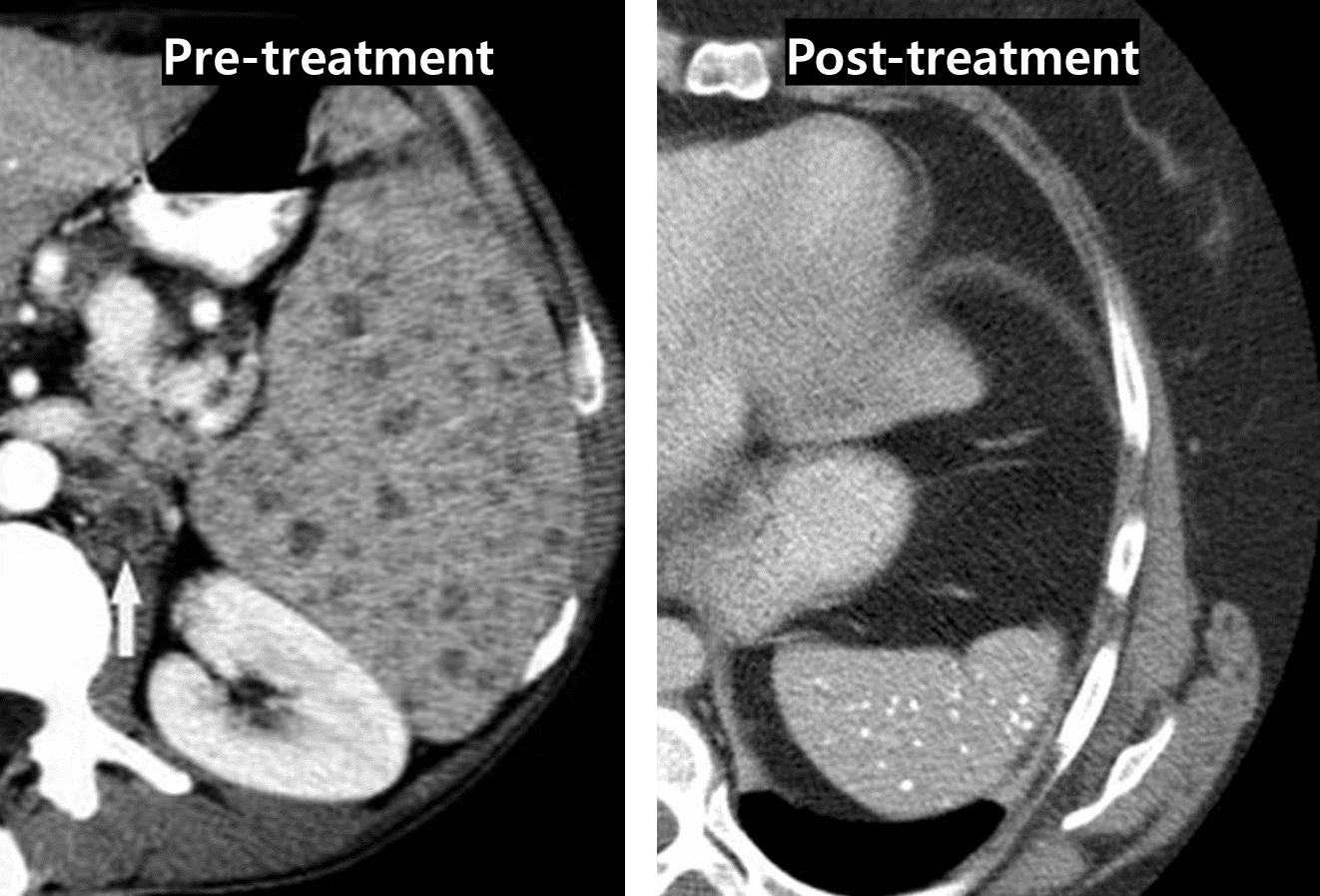
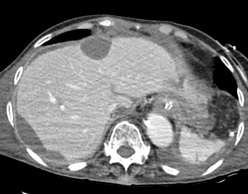
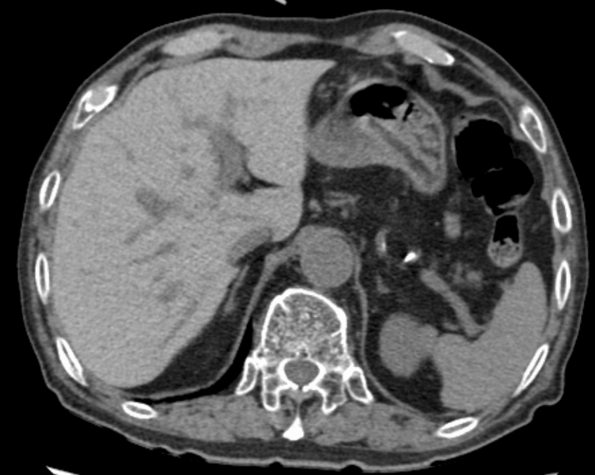
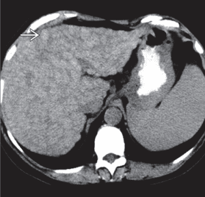
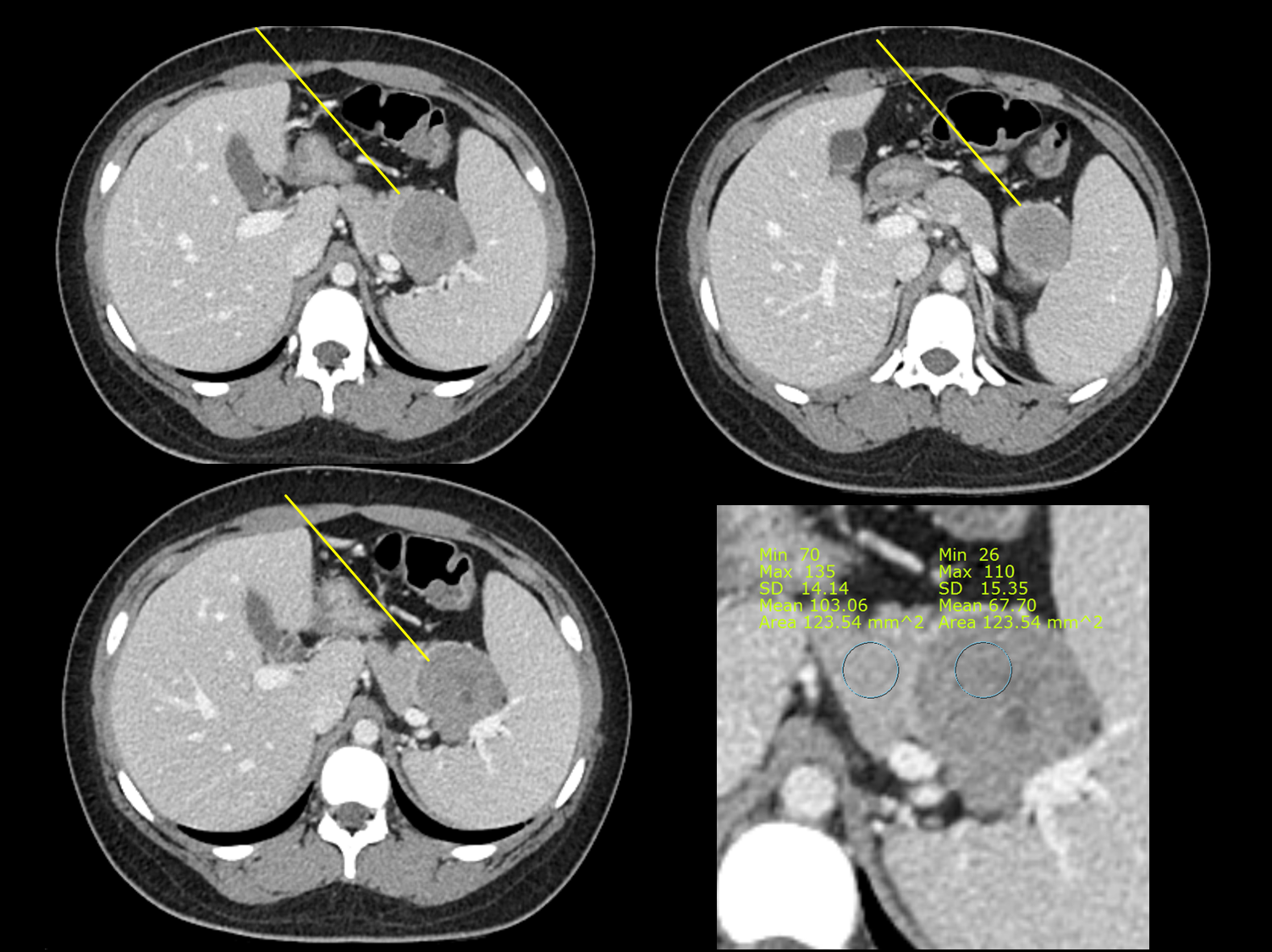
• Pyogenic abscess on CECT – Low-attenuation complex fluid collection ± air-fluid levels – Internal gas bubbles, which although uncommon, are very specific for splenic abscess – Multiloculated appearance seen with liver abscesses possible, but less common with splenic abscess – May extend to subcapsular location and may rarely cause splenic rupture with generalized peritonitis … Read more

Ischemic Bile Duct Injury radiology

Ischemic bile duct injury refers to ischemic injury of the intrahepatic bile ducts epithelium commonly seen as a complication of hepatic artery thrombosis following liver transplantation or TACE. The intrahepatic biliary ducts are exclusively supplied by the peribiliary plexus, which branches from the hepatic artery IMAGING – Biliary infarct will manifest as wall irregularities and … Read more

Pseudomyxoma Peritonei radiology

IMAGING• CT: Low-attenuation masses (usually < 20 HU) scattered throughout peritoneum – Frequently associated with loculated ascites of similar attenuation to individual implants – Implants cause mass effect on liver and spleen, producing characteristic “scalloped” appearance – Implants may demonstrate curvilinear calcification – Dominant cystic or solid mass often present in right lower quadrant (in … Read more

Hepatic hemosiderosis : CT finding

DEFINITION OF Hemochromatosis Iron overload disorder in which the involved organs store too much iron. Hemochromatosis: Classified into 2 types – Primary (hereditary): Autosomal recessive – Secondary: Due to increased iron intake, transfusions,etc DEFINITION OF Hemosiderosis – Increased iron deposition in involved organ without damage IMAGING – Liver that is hyperdense on NECT and markedly … Read more

Hepatic siderotic nodule radiology

Siderotic regenerative nodules: Paramagnetic effect of iron within nodules – Hyperdense on NECT, isodense on CECT – Hypointense on T2 and GRE MR – 3-10 mm in size Dysplastic regenerative nodules – T1WI: Hyperintense; T2WI: Hypointense – Minimal vascularity – Take up and retain hepatobiliary MR contrast agents on delayed phase – Most specific test … Read more

Mucinous adenocarcinoma : T2 hyperintense in Rectal MRI

Diagnosis of mucinous carcinoma at initial biopsy is prone to sampling error, with mis-classification in approximately 25% of cases. However, this tumor subtype has a classic appearance at MRI, allowing the radiologist to diagnose it confidently and accurately in almost all cases. Extracellular mucin is T2 hyperintense; thus, mucinous carcinomas have significantly higher T2-weighted signal … Read more

What is the diffrenece between two malignancies of stomach ?: Gastric lymphoma versus gastric adenocarcinoma

A 35 year-old male came to E.R due to melena lasting for 3 days Considering the imaging finding of circumferential wall thickening of gastric body with no sign of obstruction The first impression was gastric lymphoma But, If the lesion is lymphoma, the absence of lymphadenopathy is not common. Endoscopic biopsy was done, the pathologic … Read more

Solid pseudopapillary tumor, F/14, incidental found mass on APCT

Female / 14, Abdominal pain with diarrhea (2DA) Differential diagnosis should include solid pseudopapillary tumor(SPT), pancreatoblastoma, islet cell tumor (insulinoma) Firstly, pancreatoblastoma is frequently occured in boys less than 10 year-old and insulinoma is usually found in head of pancreas and shows vivid enhancement than normal pancreas parenchyma solid pseudopapillary tumor is more likely occurs … Read more

Focal fatty sparing of the liver: Well known mimicker of malignancy

Male / 67 years old, Incidental finding on medical screening Focal hypoechoic area just above of the IVC (collapsed due to compression of the probe) This is called ‘Focal fatty sparing of the liver’ This lesion is well known mimicker of malignancy such as metastasis or HCC The main cause of this lesions are aberrant … Read more