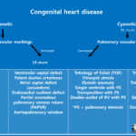
What is the Tetralogy of Fallot?
Tetralogy of Fallot (TOF) is the most common cause of
cyanotic congenital heart disease, accounting for approximately 10% of congenital cardiac malformations.
Cause of tetralogy of Fallot
Tetralogy of Fallot is resulted from underdevelopment of the subpulmonary infundibulum(Conus).
– Anterosuperior deviation and hypertrophy of conal septum
Tetralogy of Fallot is characterized classic four findings :
(a) Ventricular septal defect (VSD)
(b) Overriding aorta : Aorta is positioned directaly over a VSD instead of over the ventricle
(c) Right ventricular outflow tract obstruction (RVOTO)
(d) Right ventricular hypertrophy (RVH)
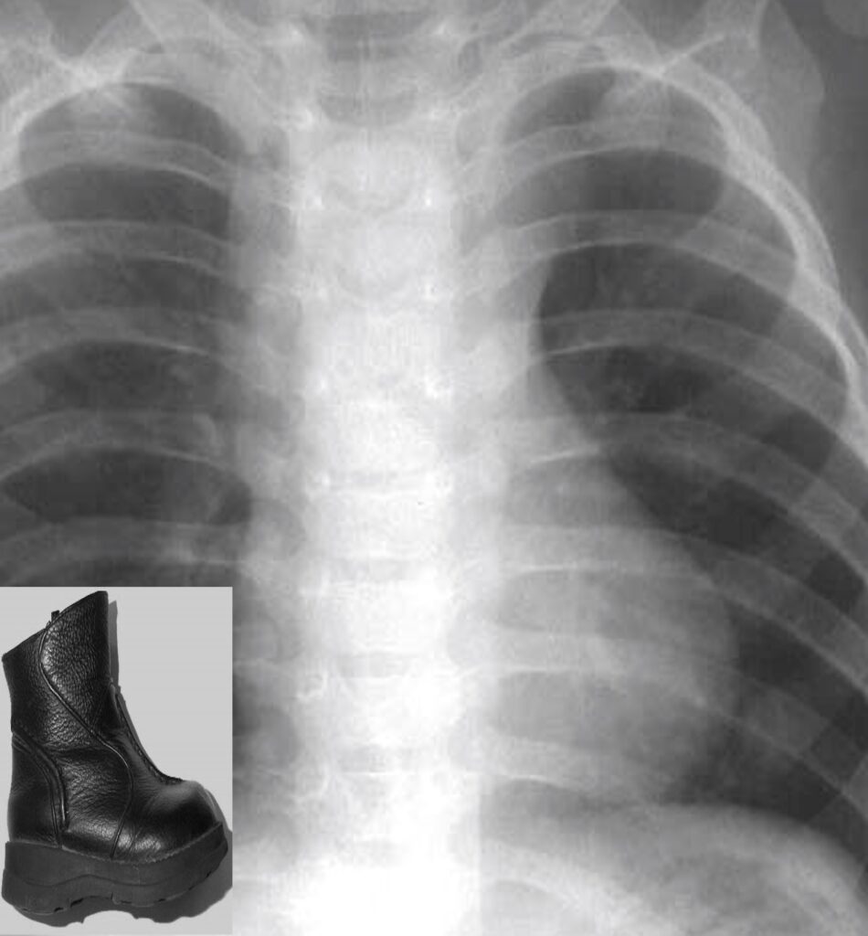
Imaging findings of tetralogy of Fallot
Boots shape
– RVH
– Round apex
– Elevated LV
– Clockwise rotation of the heart
Treatment of Tetralogy of Fallot
(a) Complete primary repair, which is usually undertaken within the first year of life
– Closing the VSD, relieving the RVOTO
(b) Surgical palliation with placement of Blalock-Taussig shunt (BT shunt) to augment pulmonary perfusion may be performed when the central pulmonary artery anatomy is inadequate
– Connecting subclavian or innominate artery with ipsilateral pulmonary artery
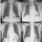
Note that the pulmonary vascular markings are decreased on chest radiograph.
This MAPCA surrounds the bronchus intermedius (white arrow); its origin from the descending thoracic aorta is not shown.

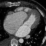
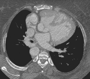
Black arrow : VSD
White arrow : overriding aorta
with hypertrophied right ventricle
Reference)
Charles S. White, Linda B. Haramati, Joseph Jen-Sho Chen, and Jeffrey M. Levsky (2014), Cardiac Imaging, Oxford university press