1. Azygos vein
Reflect pressure of right atrium
2. Vascular pedicle width
– Reflect total body blood flow
– The vascular pedicle width (VPW) is the distance between parallel lines drawn from the point at which the SVC intersects the right main bronchus an a line drawn at the takeoff of the left subclavian artery from the aorta.
– The mean vascular pedicle width is 38-58 mm on posteroanterior chest radiographs.
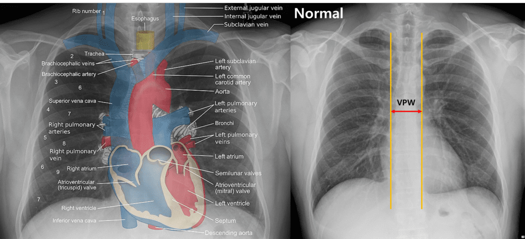
Right) Vascular pedicle width on chest x-ray
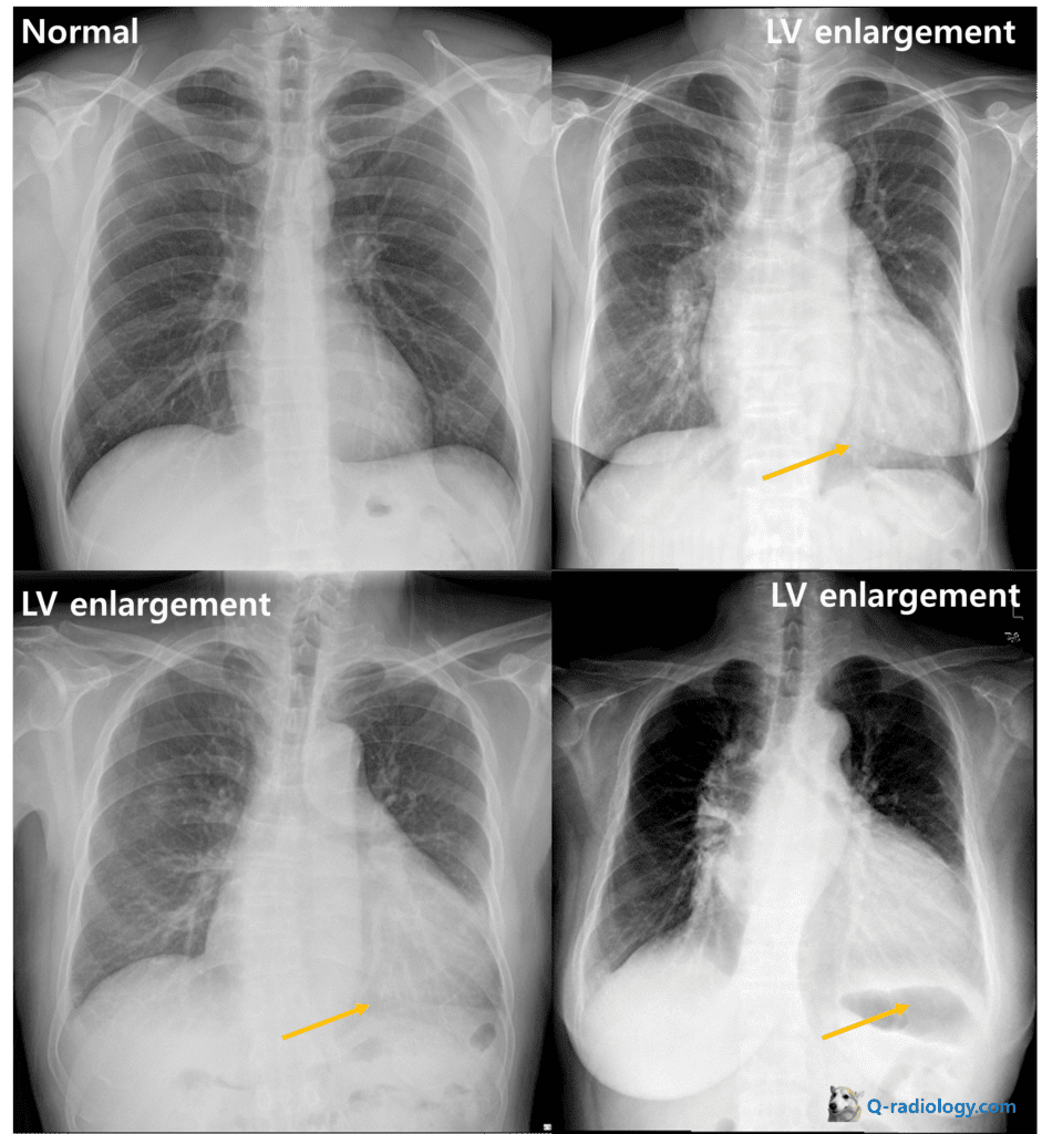
3. Left ventricular enlargement (LVE)
Left lower downward migration of apex (towards below the left side diaphragm)
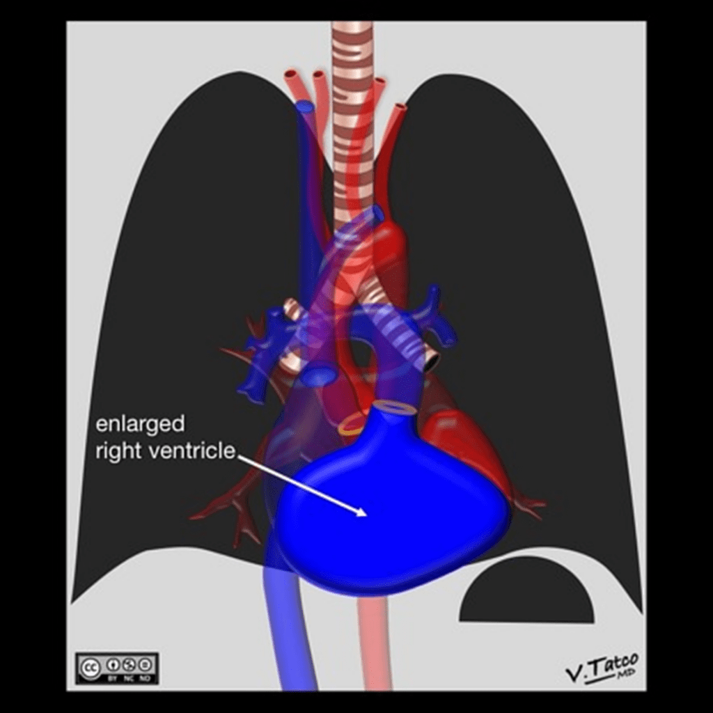
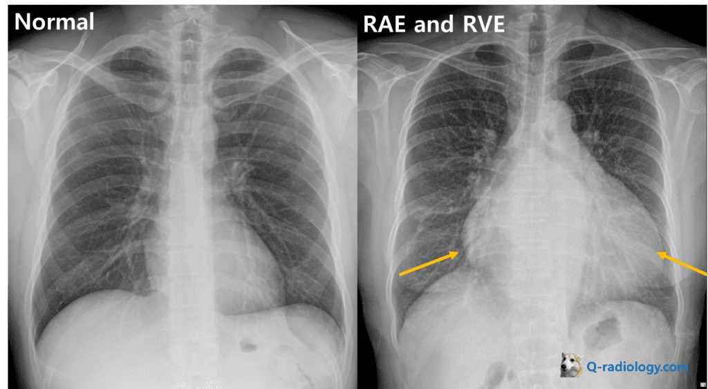
4. Right ventricular enlargement
– left and upward migration of apex
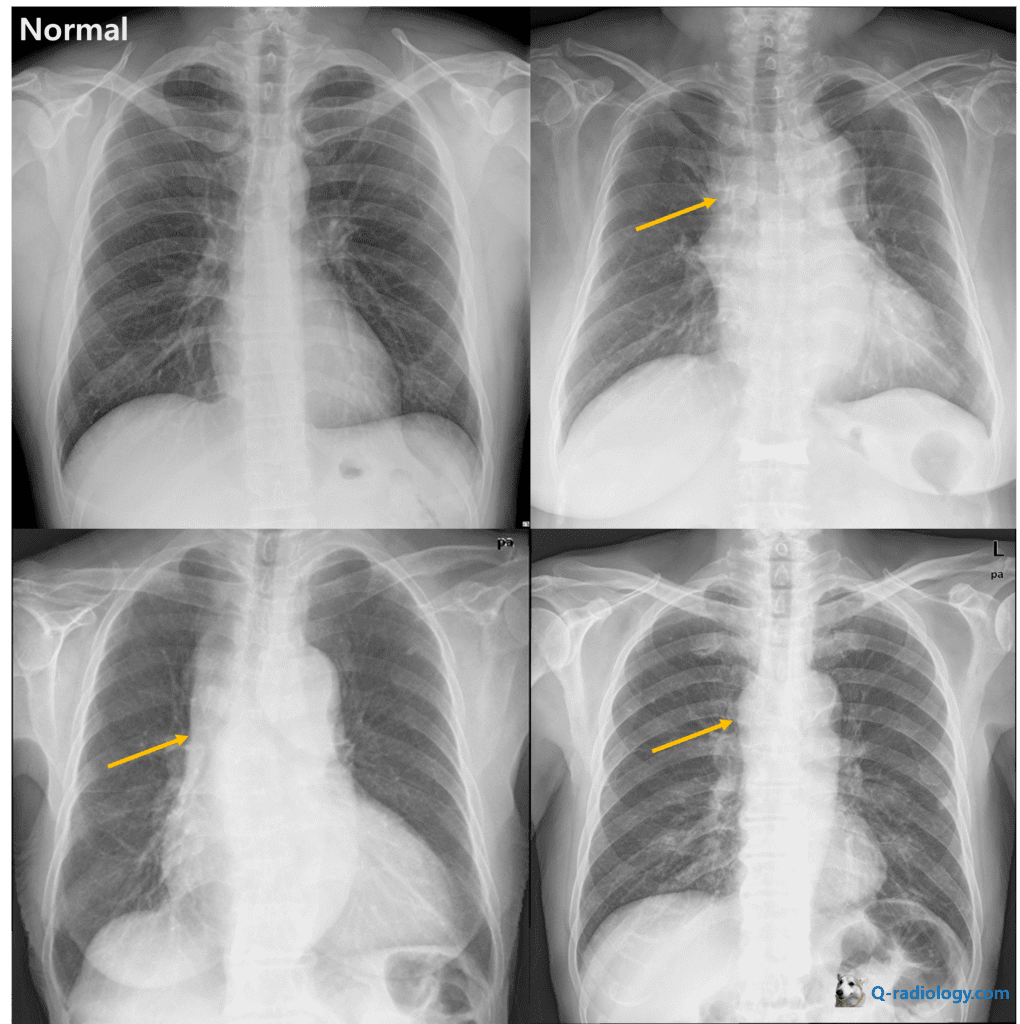
5. Left atrial enlargement
– Double contour sign
– Uplifted left main bronchus
– Enlarged left atrial appendage

Note that contrast reversal makes double contour sign more clear
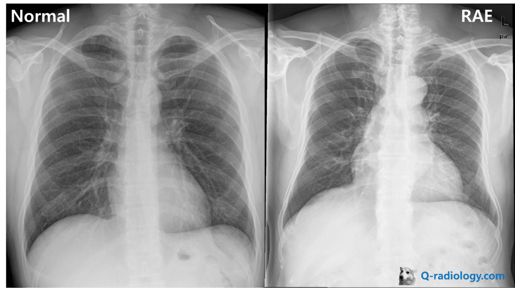
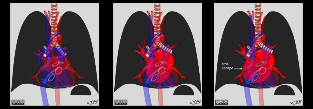
6. Right atrial enlargement
– Lower margin of right cardiac border is more convex than normal