Solitary bone plasmacytoma: Back pain without trauma history, could be sign of bone tumor

An 82-year old female presented E.R with severe back pain

Solitary bone plasmacytoma
Arrow : wedge shape T6 vertebra, suggesting compression fracture

Solitary bone plasmacytoma
The posterior border of T6 shows bulging contour with soft tissue density occupying vertebral body. Cortical strut thickening is also found (Mini brain sign). These findings are suggesting pathologic fracture rather than acute compression fracture.

Solitary bone plasmacytoma
The osteolytic lesion shows Iso signal intensity to marrow on T1WI. after injecting contrast materials, the lesion shows homogenous enhancement. The lesion is surrounded low signal intensity rim (corresponding with the sclerotic rims on CT ; mini brain sign).

Solitary bone plasmacytoma
The mass extended to paravertebral space. shows expansile pattern rather than infiltrative one.

Solitary bone plasmacytoma
PET CT) Hypermetabolic lesion at T6 and stomach.

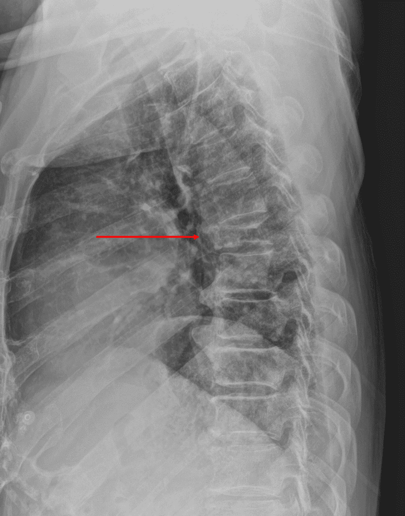
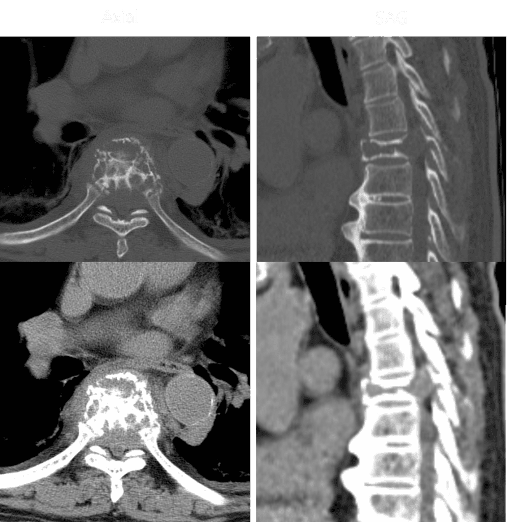
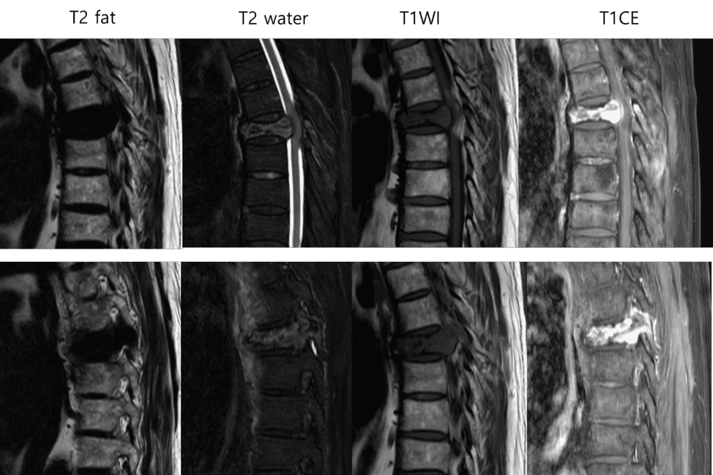
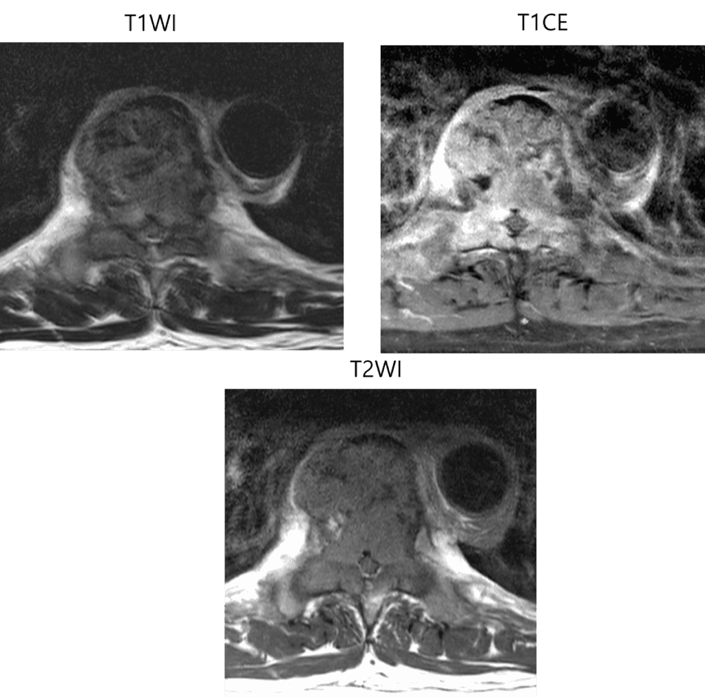
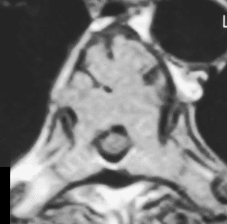
The most possible diagnosis was plasmacytoma, considering CT and MRI findings.

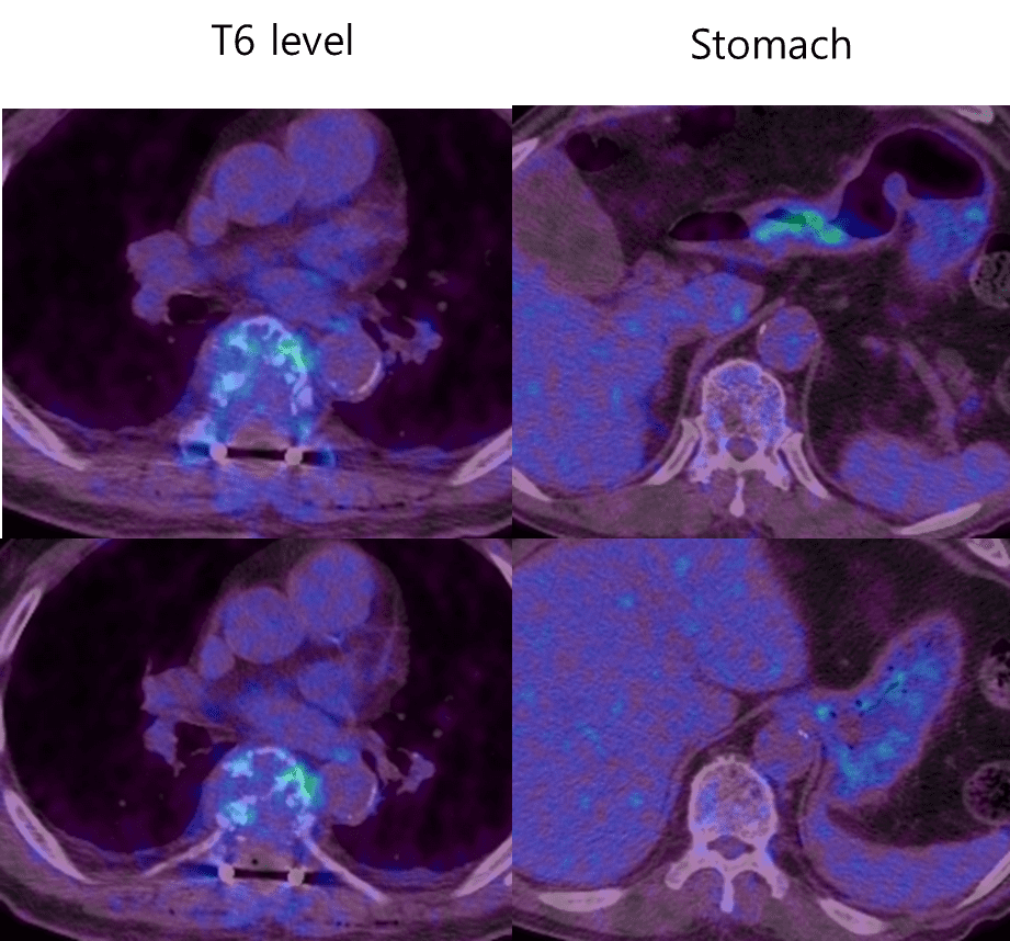
The PET-CT report suggest the possibility of stomach cancer and its bone metastasis.

Final diagnosis : Spine (T6), excision

-> Plasmacytoma

    Note :

     Results of immunohistochemical stainings:

        cytokeratin,  (-);  leukocyte common antigen (-), (0); CD99,  (-);

        Fli-1,  (0);  CD31,  (-);  TLE-1,   (+);   Ki-67  labeling index, (5%).

Solitary bone plasmacytoma

Solitary monoclonal plasma cell tumor of bone or soft tissue, without evidence of multiple myeloma (MM)

Location

  • Axial skeleton (m/c), followed by extremities
  • Thoracic > Lumbar > Cervical
  • Rib, sternum, clavicle, scapula ; 20% of all cases

Demographics

  • Age : mean age – 55yrs
  • M > F (2:1)
  • 3-5% present monoclonal gammopathy

Imaging finding

  • CT
    • Lytic, compression fracture +/- soft tissue mass
  • MRI
    • Marrow iso to hypo signal intensity (T1WI)
    • Mini brain
    • Posterior element involvement (common)
    • Draped curtain sign
    • Heterogeneous signal intensity on T2WI
    • Mild to moderate diffuse enhancement
The “mini brain” ; axial T2WI MR image reveals diffuse high SI throughout vertebral body and low SI cortical struts resembling mini brain AJR 2000;175:261–263

References)

Diagnostic Imaing, Spine 2nd edition  p. 5-64 ~ 67

AJNR AM J Neuroradiol. 2010 Mar;31(3):559-64

AJR 2000;175:261–263

0 0 votes
Article Rating
Subscribe
Notify of
guest
0 Comments
Inline Feedbacks
View all comments
0
Would love your thoughts, please comment.x
()
x