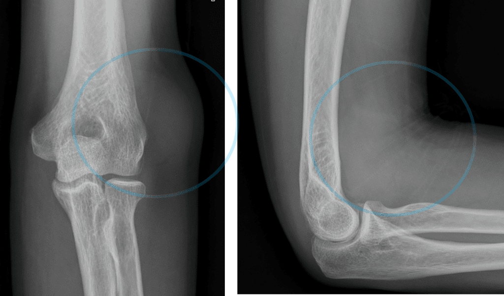
1) Well defined high density mass like attenuation at lateral aspect of left distal humerus, without associated bony erosion nor sclerosis
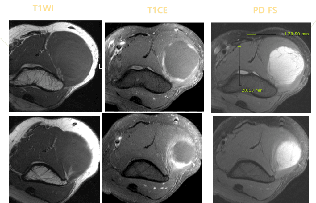
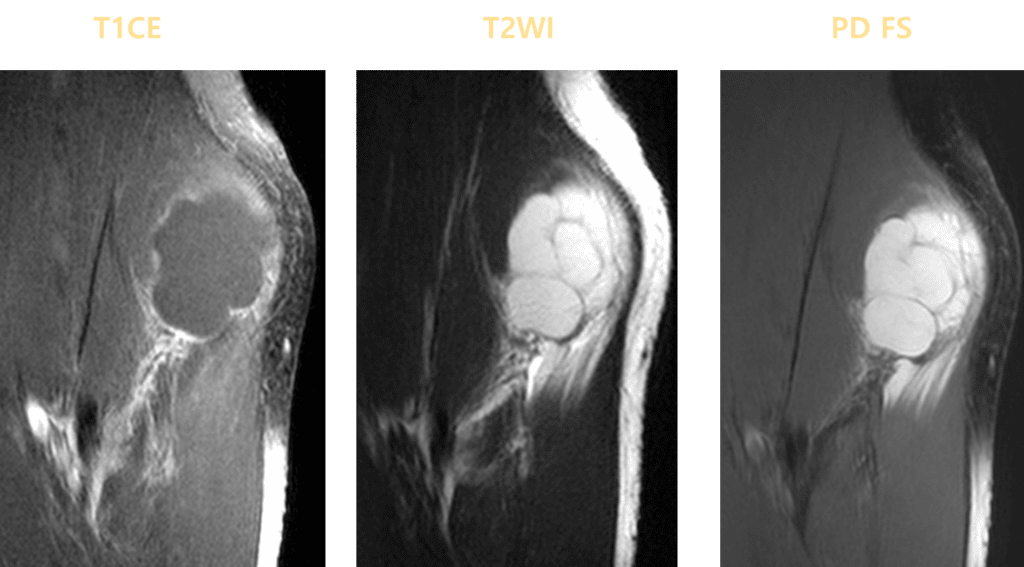
- T2 hyperintense, T1 hypointense mass with perilesional fat rind
Differential Diagnosis: benign peripheral nerve sheath tumor, ganglion, cyst, other myxoid neoplasms
Biopsy confirmed Intramuscular myxoma
Intramuscular myxoma
is the most common benign myxoid neoplasm, consisting of abundant myxoid stroma interspersed with benign spindle cells.
Intramuscular myxoma is most frequently diagnosed in patients 40–70 years of age.
The most common presentation is a slowly growing painless mass.
Intramuscular myxomas are typically solitary.
When multiple myxomas are present, they are almost always associated with fibrous dysplasia, known as Mazabraud syndrome.
Most musculoskeletal myxomas are intramuscular (82%) in location, occurring most often in the thigh, followed by upper arm, calf, and buttock.
On MRI, the lesions are well circumscribed with smooth or slightly lobulated margins, homogeneously hypointense on T1-weighted images, and extremely hyperintense on T2-weighted images.
On contrast-enhanced images, lesions show variable heterogeneous, sometimes avid, contrast enhancement due to the presence of abundant vascularity within the myxoid matrix.
The presence of a perilesional rind of fat or perilesional edema has been described to be highly suggestive of the diagnosis on MRI.
Surgical excision with wide margins is the treatment of choice for intramuscular myxomas, with recurrence being rare.
References:
1. Kransdorf MJ, Murphey MD (2014) Imaging of soft tissue masses In:
Kransdorf MJ, Murphey MD. Imaging of soft tissue tumors. 3rd ed. Philadelphia,
Pa: Lippincott Williams & Williams
2. Petscavage-Thomas JM, Walker
EA, Logie CI, Clarke LE, Duryea DM, Murphey MD. Soft-tissue myxomatous lesions:
review of salient imaging features with pathologic comparison. Radiographics
2014;34:964-80
3.Baheti AD, Tirumani SH, Rosenthal MH, et al. Myxoid soft-tissue neoplasms: comprehensive update
of the taxonomy and MRI features. AJR Am J Roentgenol. 2015;204:374-85
