Krukenberg tumor: Bilateral complex ovarian masses with high signal intensity on T2 weighted image 2022-11-05 by Q-radiology
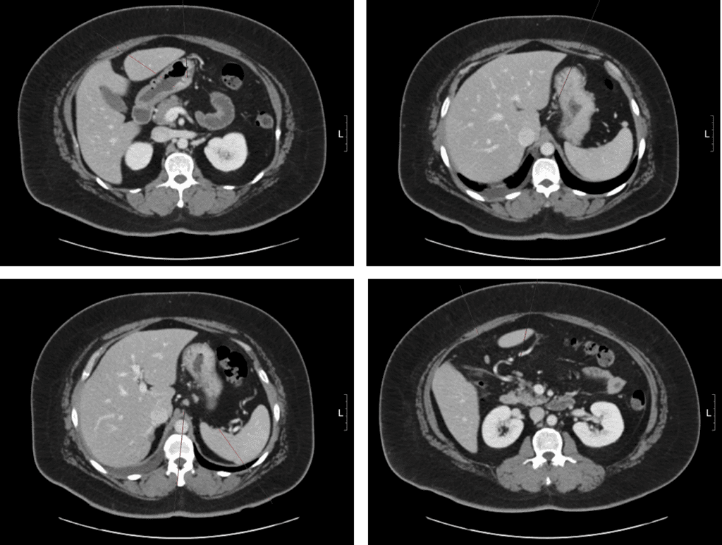
A 44 year-old female who had been diagnosed advanced gastric cancer had CT and PET-CT scan for metastases work upStomach, lesser curvature of low body to angle, endoscopic biopsy; 
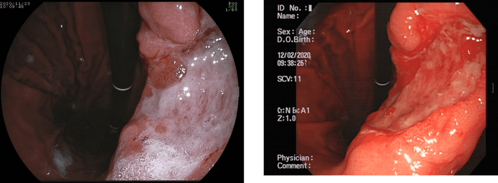
POORLY COHESIVE CARCINOMA Focal enhancing wall thickening with ulceration at gastric body with small to borderline lymphadenopathy at left gastric artery, right gastriepiploic artery, and small bowel mesentery. Homogenously enhancing masses at bilateral ovaries are found in the same scan, metastasis cannot be excluded.PET-CT) Hypermetabolic wall thickening at the gastric angle (SUVmax= 3.4).
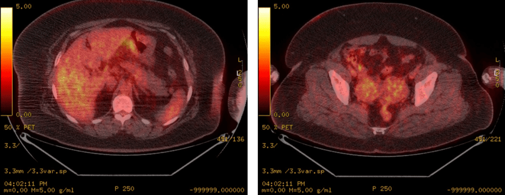
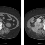
Mild hypermetabolic masses in the both adnexa (SUVmax= 3.8).Pelvis MRI taken after two months of neoadjuvant chemotherapy.
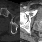
Heterogeneously enhancing complex solid masses are found in both ovaries. the bilaterality suggests that the lesions are metastasis.T2WI reveals high signal intensity cystic portion of the tumors, suggesting mucin component.These imaging findings are match for the Krukenberg tumor Reference) RadioGraphics Nov 1 2002; 1305–1325Related posts: Prostate abscess, fever for 1 week, 82 years old male patient What is Malacoplakia