DEFINITION
Autoimmune disease characterized by recurrent inflammation of cartilaginous structures
CLINICAL FINDING
– Common in 20~30s, but all age group are possible
– Auricular chondritis
– Ocular manifestations (scleritis, episcleritis, or conjunctivitis)
– Nasal chondritis (painful inflammation of nasal cartilage)
– Costochondritis (retrosternal chest pain)
– Laryngotracheal and pulmonary involvement (50% during disease course)
– Respiratory complications and lower respiratory tract infections represent most common causes of death
IMAGING
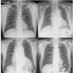
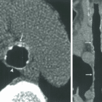
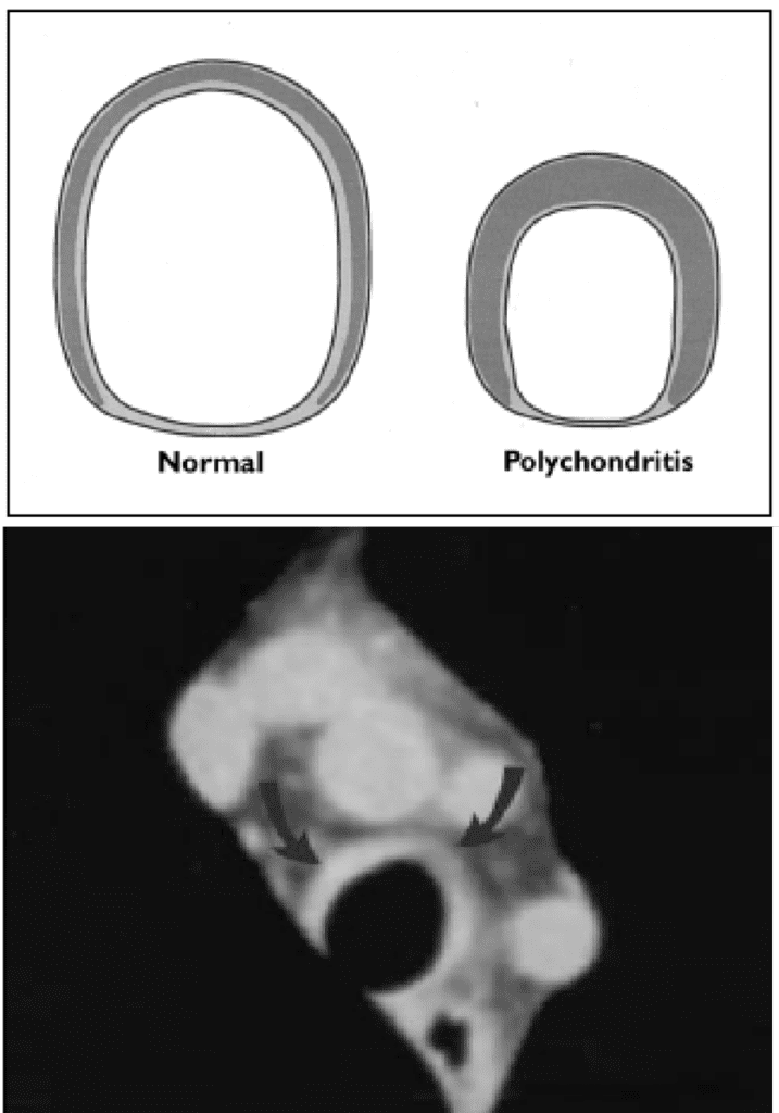
– Diffuse tracheobronchial narrowing (late finding) with smooth wall thickening
; wall thickness > 2mm with/without calcification
; sparing posterior membranous trachea
– Increased airway well attenuation
– Destruction of cartilaginous rings
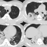
– Excessive collapsibility, often associated with air-trapping (expiratory CT)
– Mosaic attenuation
– Obstructive bronchiectasis
DIFFERENTIAL DIAGNOSIS
– TBO : multiple nodules in tracheal wall sparing posterior membranous trachea
– Amyloidosis : irregular wall thickening, not sparing the posterior wall
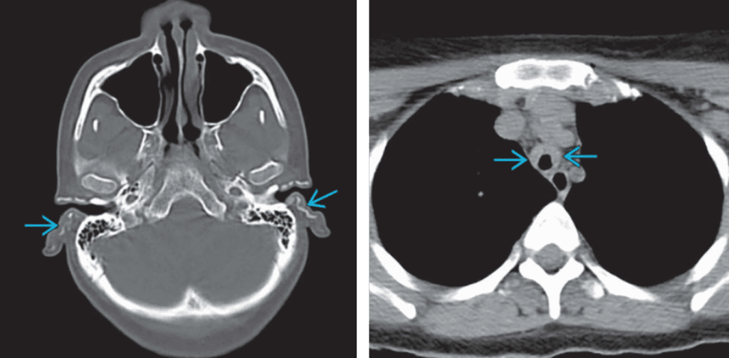
RIght) Relapsing polychondritis involving trachea, marked smooth wall thickening of trachea is seen