Male / 44 , Chief complaint : fever (since 6 days ago)
Never smoker
The patient’s main symptom is fever continued for 6 days.
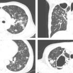
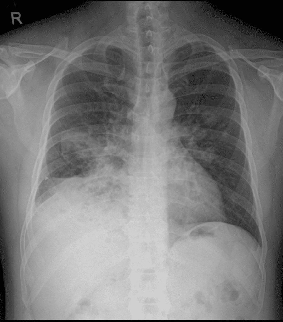
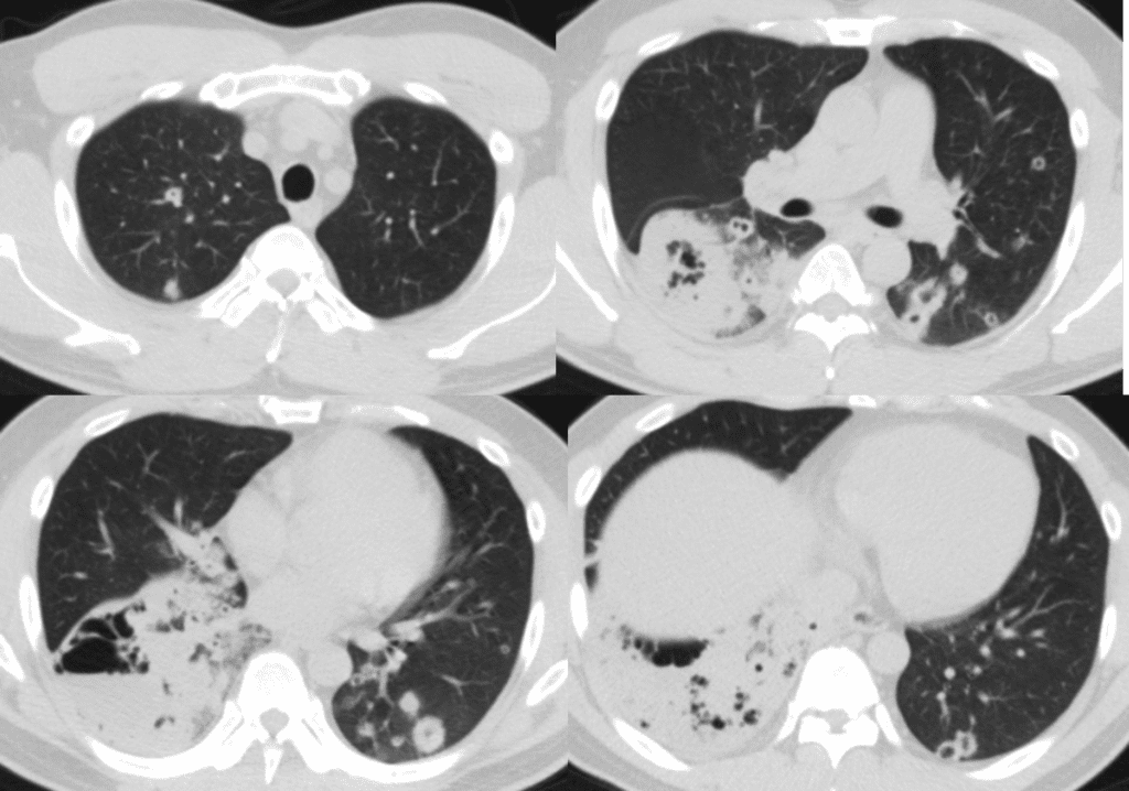
the chest radiograph and CT scan seems to be lobar pneumonia.
the patient was treated with antibiotics for 5 months
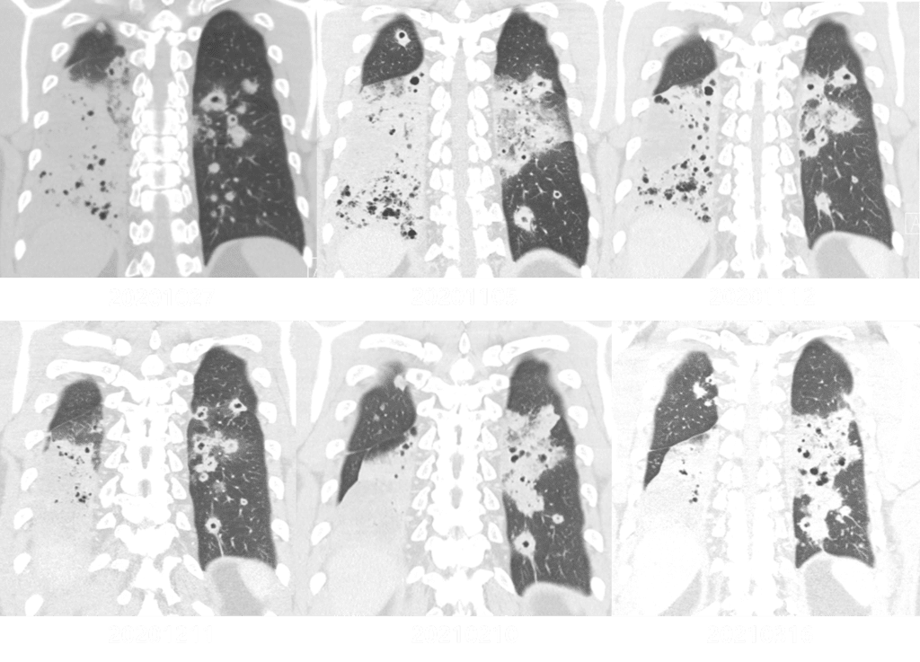
Long standing lobar consolidation made us to suspect the malignancy especially, invasive mucinous adenocarcinoma with metastasis
we discussed the possibility of vasculitis such as granulomatous polyangiitis but distribution of the lesion is not explainable (in GPA it would show more diffuse distribution)
So surgical biopsy was planned for diagnosis
wedge resection was done, the result was
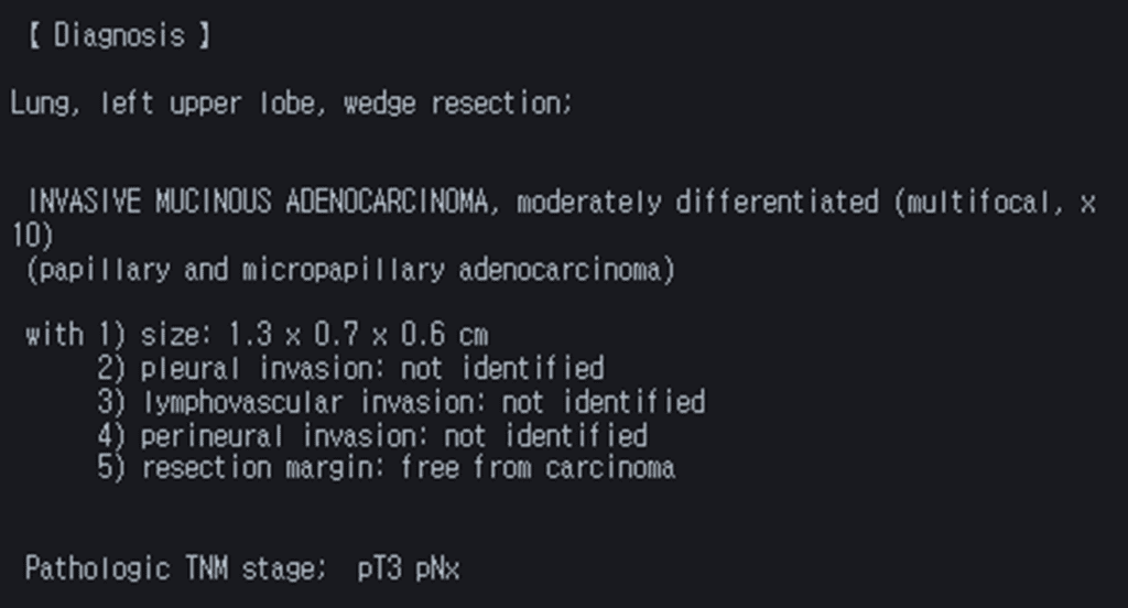
Invasive mucinous adenocarcinoma is variants of invasive adenocarcinoma in IASLC (International Association for the Study of Lung Cancer) classification and former mucinous BAC (bronchoalveolar carcinoma)
goblet or columnar tumor cells with abundant intracytoplasmic mucin are typically shown in pathology of invasive mucinous adenocarcinoma
Take home message : invasive mucinous adenocacinoma should be suspected long standing (over 6 weeks) lobar consolidation (no or less improvement with antibiotics)
Reference)
Transl Lung Cancer Res . 2017 Oct;6(5):508-512.
J Thorac Oncol . 2011 Feb;6(2):244-85.
송재우, 이창현, 구진모, 서준범. (2015). 흉부영상의학. 2nd ed. 일조각.
Thoracic Cancer, Volume: 11, Issue: 12, Pages: 3463-3472