1. What is myocarditis?
Myocarditis is inflammation of the myocardium, most often secondary to a viral infection involving the upper respiratory or gastrointestinal tract.
Other less common etiologies include adverse drug reactions and autoimmune phenomena (e.g., sarcoidosis, systemic lupus erythematosis).
As definitive diagnosis requires tissue biopsy, the true incidence of the disease is unknown and thought to be underreported.
2. Clinical features
– Flu-like symptom, Frequently elevated cardiac enzyme
– Difficult to differentiate with acute myocardial infarction by clinical symptoms
– Mostly self limited, but can progress to DCMP
3. Imaging features
MRI
– T2WI : edema
– Early enhanced T1WI : hyperemia and capillary leak
– DE-MRI : multiple patchy enhancement predominantly in subepicardial portion
– Not related to vascular territory
– Useful for the guidance of tissue biopsy (enhancing portion)
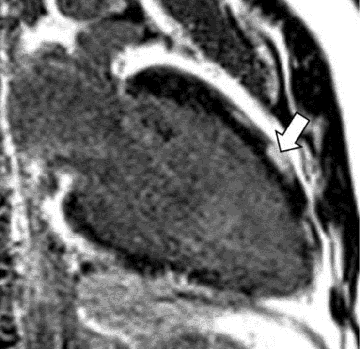
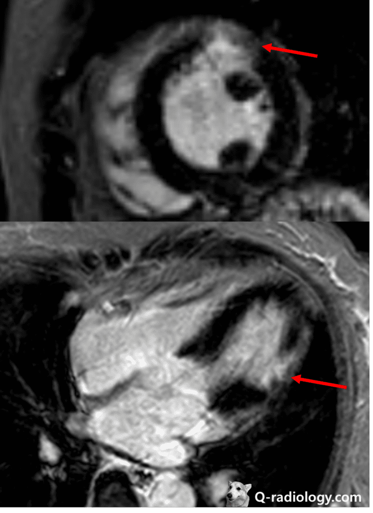
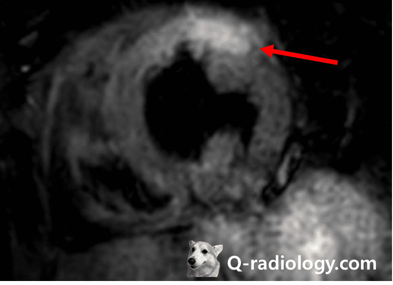
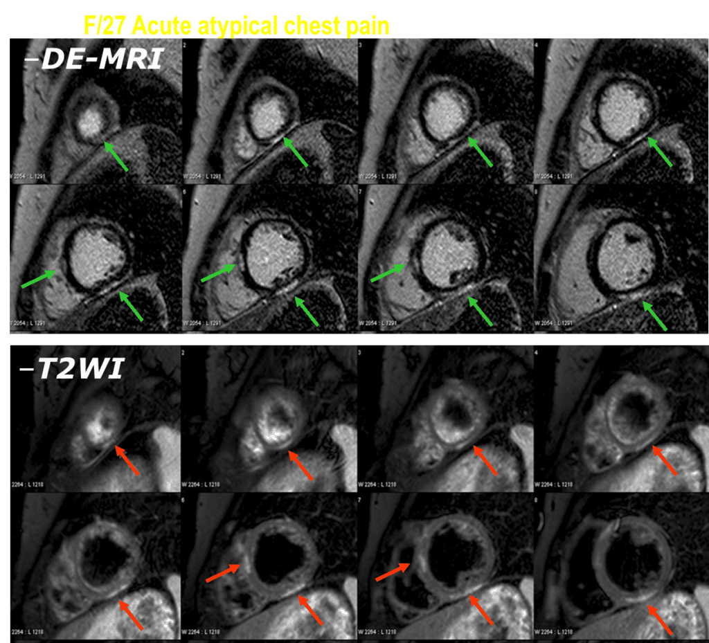
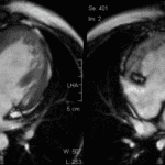
Enhancement pattern is non-vascular territory and non-subendocardial dominant area.
T2-weighted MRI show ill-defined high signal area at same area (arrows).