Mitral valvular stenosis
What is mitral stenosis?
Mitral stenosis (MS) is characterized by narrowing of the valve orifice compromising blood flow between the left atrium and ventricle.
What is the causes of mitral stenosis?
By far the most common etiology of MS is rheumatic heart disease.
Less common causes are infective endocarditis, severe mitral annular calcification, congenital pathology, and obstructive lesions, including atrial myxoma, large vegetations, or thrombi.
Epidemiology of mitral stenosis
With earlier recognition and awareness of streptococcal infections, the incidence has decreased dramatically in the United States to roughly 1 case per 100,000.
In developing nations, however, rates can approach 150 per 100,000. The female to male ratio is 2:1, with the age of onset usually occurring in the third or fourth decade.
Imaging finding of mitral stenosis
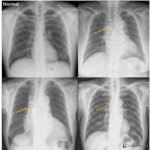
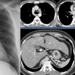
Enlarged left atrium with or without wall calcification
– double density sign (LAE)
– bulge of superior posterior cardiac border below carina
– displaced esophagus, Right posteriorly
– left main bronchus elevation
– dilated left atrial appendage
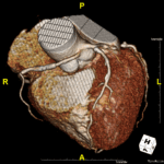
Calcification of valve leaflet
Prominent PA segment
Small aorta
RVH, RVE
Pulmonary edema
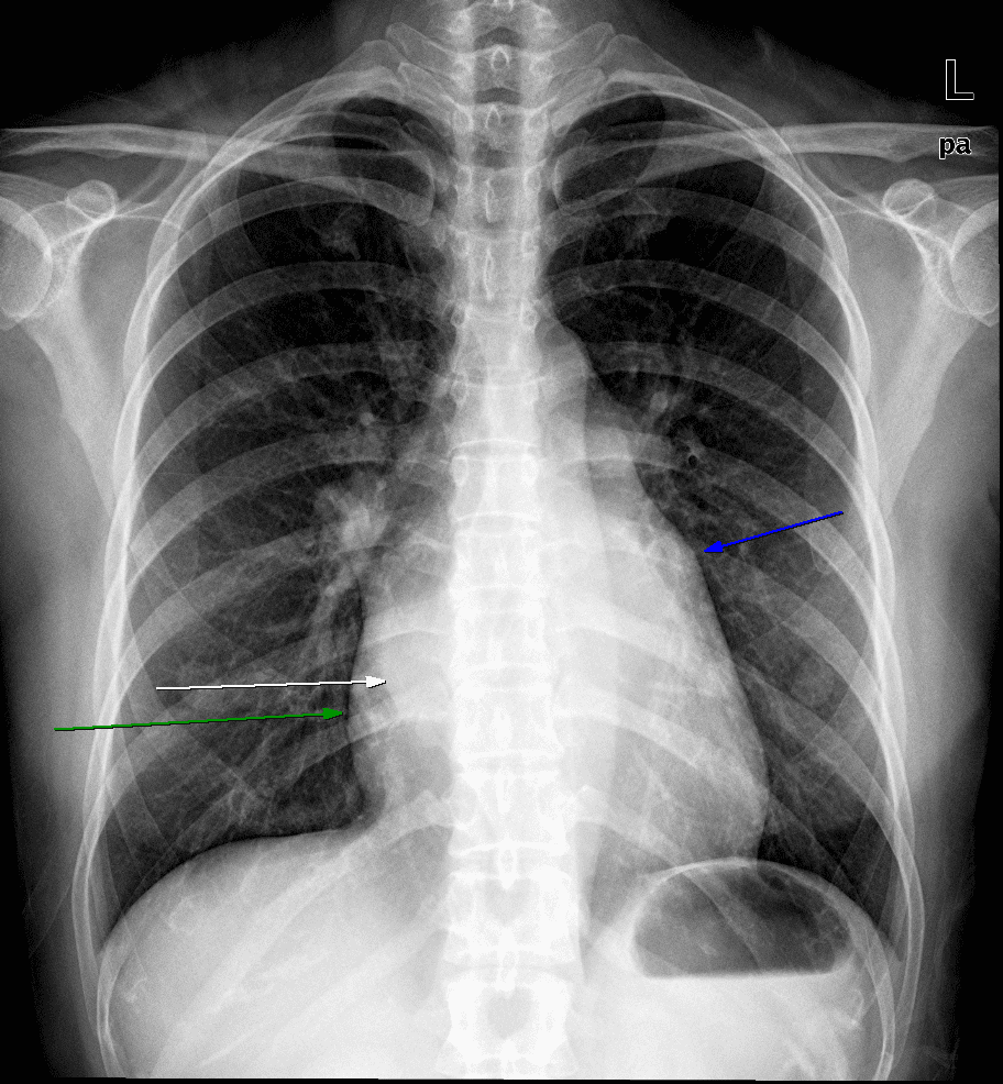
White and green arrows : Double density sign, indicating left atrial enlargement.
Blue arrow : Dilated left atrial appendage
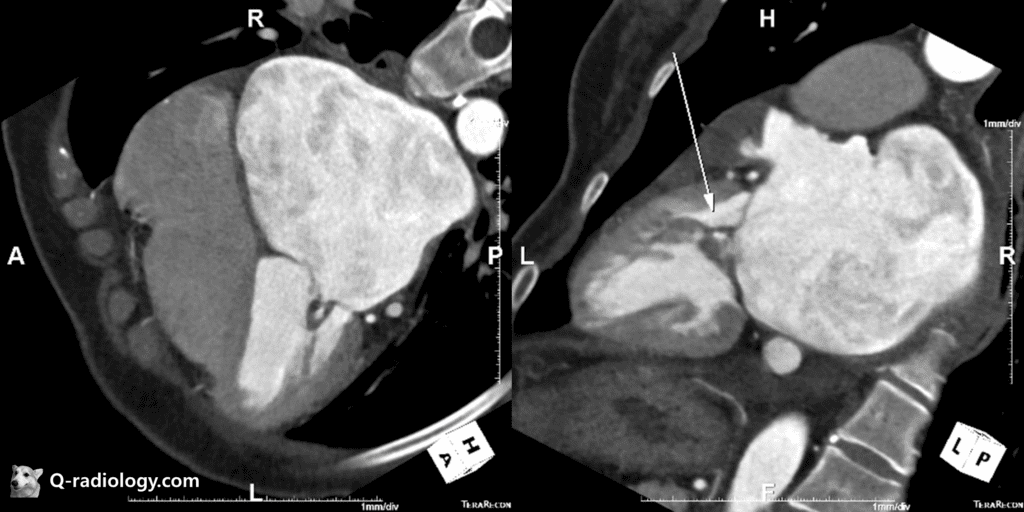
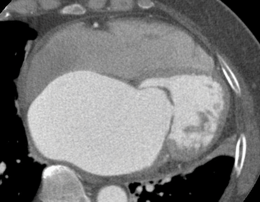
Mitral valvular thickening with calcification.
Left atrial enlargement.
Mitral valve bowing toward left ventricle with left atrial enlargement.
Pathophysiology
– LA, LV pressure increase
– PVP : >25 mmHg, pulmonary edema
Normal mitral valve orifice : 4-6 cm2
> 1.5 cm2 : mild MS
1.0cm2 – 1.5cm2 : moderate MS
< 1cm2 : severe MS
Complications
– Infective endocarditis
– LA thrombi and emboli
See more about cardiovascular imaging
Follow my instagram

Reference)
Charles S. White, Linda B. Haramati, Joseph Jen-Sho Chen, and Jeffrey M. Levsky (2014), Cardiac Imaging, Oxford university