Noncyanotic, increased pulmonary blood flow
An 19 year-old male patient presented clinic because of an abnormal chest CT finding
What is the partial anomalous pulmonary venous return (PAPVR)?
PAPVR is a congenital anomaly that involves drainage of one to three pulmonary veins into the systemic veins, creating a partial left to right shunt.
Characteristics of PAPVR
Incidence
– 0.5% in the general population.
Associated abnormality
– Most commonly ASD, approximately 25%
Symptom
– Asymptomatic, usually incidental radiographic finding
– Lt-to-Rt shunt, pulmonary hypertension, Rt-sided heart failure
Treatment
– Surgery
– Indication
a) LR shunt ratio exceeding 2:1
b) Coexistent congenital heart disease
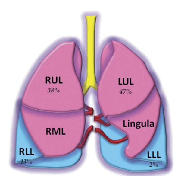
Left upper lobe, most common (47%)
Right upper lobe (38%)
Right lower lobe (13%)
Left lower lobe (2%)
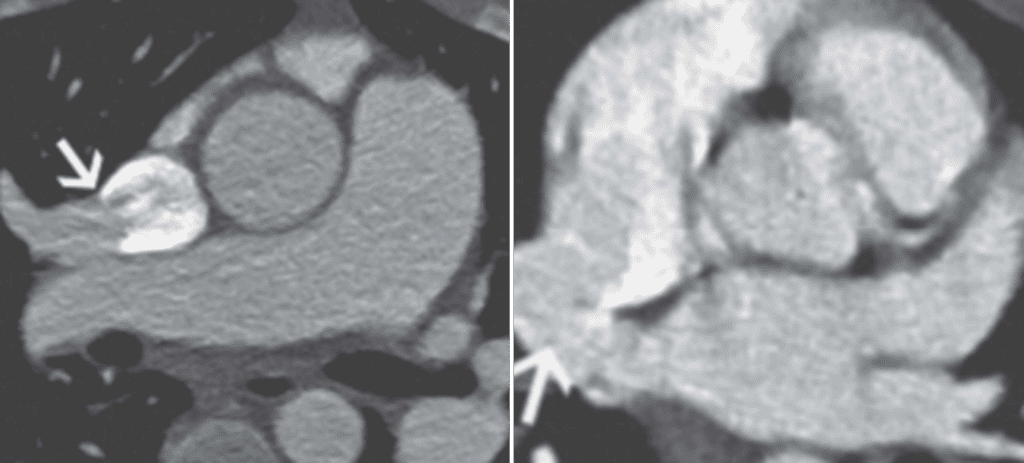
RUL PAPVR
– Drains into SVC
– Often associated with sinus venosus ASD
RLL PAPVR
– Drains into IVC
– Scimitar syndrome (Combination of pulmonary hypoplasia and partial anomalous pulmonary venous return)
– Scimitar syndrome specifically is exceedingly rare, with an incidence of just 2 per 100,000 births
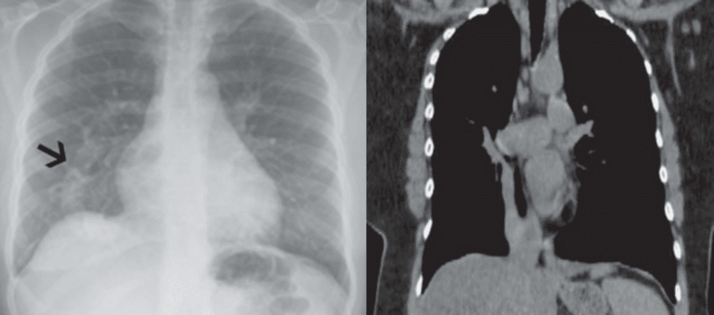
Small right hemithorax, Right lung hypoplasia
Curvilinear structure at the base of the right lung curving toward the right cardiophrenic angle, PAPVR to the IVC.
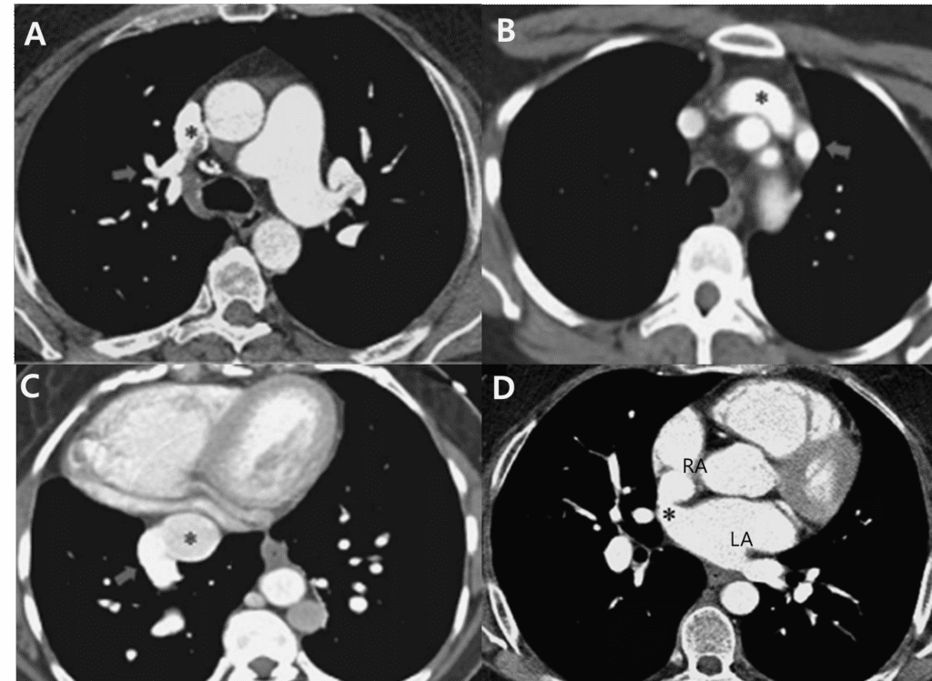
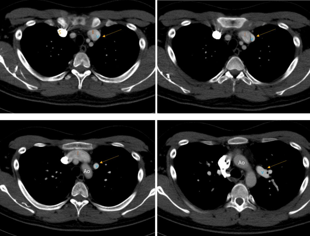
B) Left upper lobe PAPVR draining into the left innominate vein
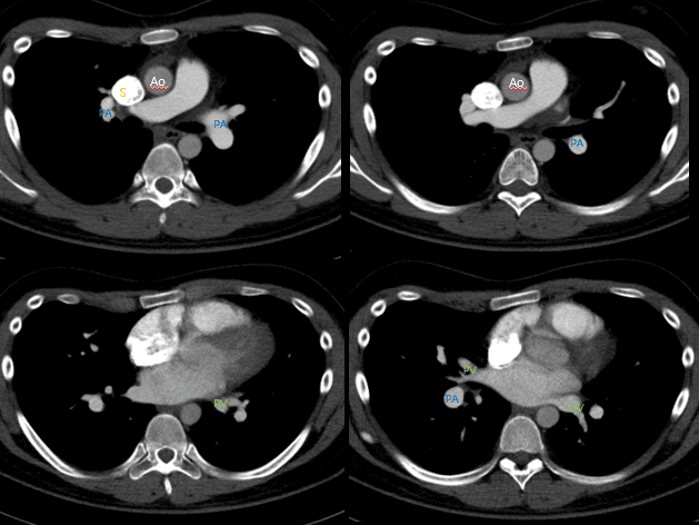
C) Right lower lobe PAPVR draining into the inferior vena cava (IVC)
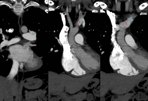
D) Sinus venosus ASD in a patient with right upper lobe PAPVR
Reference)
Charles S. White, Linda B. Haramati, Joseph Jen-Sho Chen, and Jeffrey M. Levsky (2014), Cardiac Imaging, Oxford university press
Jud W. Gurney, Helen T. Winer-Muram, et al. Diagnostic imaging Chest, II-4 6~7
Charles S. White, Linda B. Haramati, et al. Cardiac imaging.
Ho ML, Bhalla S, Bierhals A, et al. MDCT of partial anomalous pulmonary venous return (PAPVR) in adults. J Thorac Imaging 2009; 24:89–95