What is ventricular septal defect?
A ventricular septal defect (VSD) is a hole in the interventricular septum.
It is one of the most common congenital malformations of the heart, accounting for 20–30% of congenital heart defects.
VSDs may be isolated or seen associated with complex cardiac malformations, including tetrology of Fallot and transposition of the great vessels, or syndromes such as trisomy 21
Imaging finding of ventricular septal defect
Increased pulmonary vascularity (Left-to-right shunt)
Left atrial enlargement
Left ventricular enlargement
Normal or small aorta (relatively normal to ASD)
Convex pulmonary outflow tract
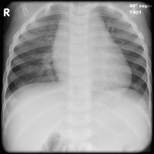
Increased pulmonary vascularity due to Left-to-right shunt
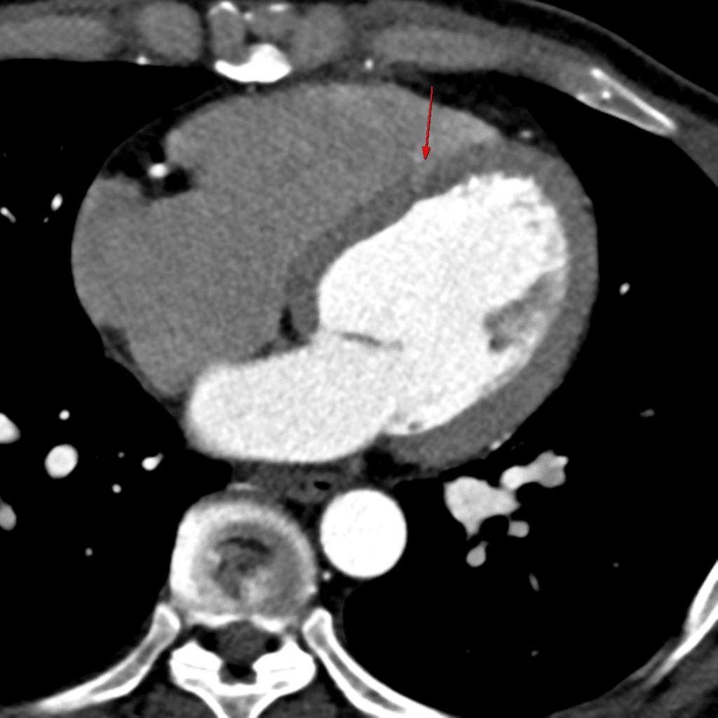
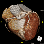
Vasalva sinus aneurysm herniated through VSD
– Associated with rupture, aortic regurgitation ➜ Consider valve replacement
Septal aneurysm = spontaneously closed VSD ➜ Observation
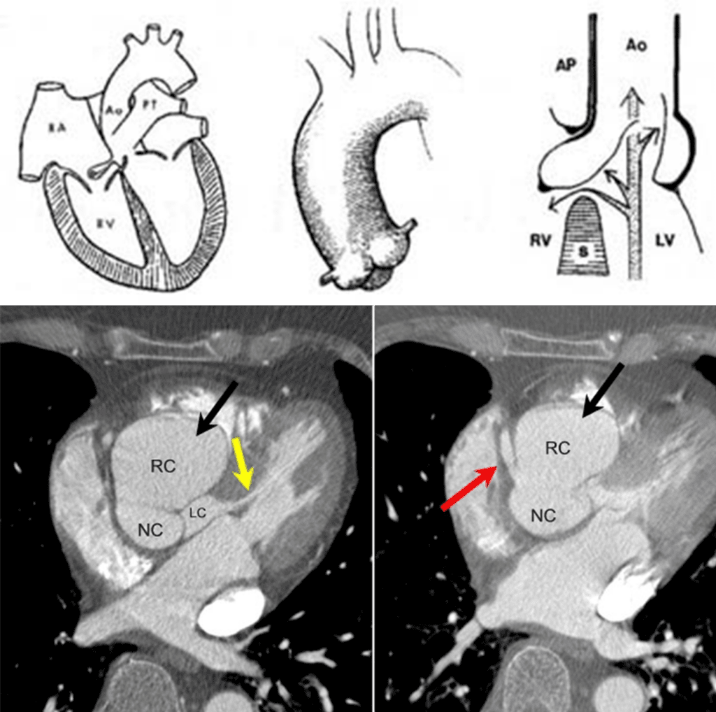
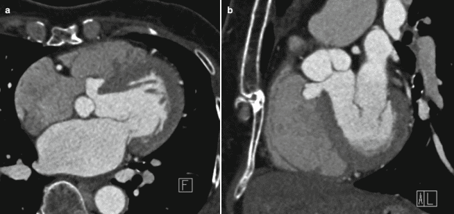
Note that aortic valves are intact.
spontaneously closed VSD
Reference)
Charles S. White, Linda B. Haramati, Joseph Jen-Sho Chen, and Jeffrey M. Levsky (2014), Cardiac Imaging, Oxford university press