DEFINITION
– Superior vena cava syndrome (also called SVC obstruction) is a group of symptoms that occurs when SVC is obstructed by intraluminal or extrinsic disease.
– SVC syndrome is characterized by Impaired venous return from head, neck, upper extremities, and trunk
CLINICAL FINDING
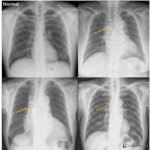
– Face, neck, upper trunk, and upper extremity edema
IMAGING
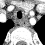
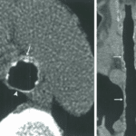
CT and MR
– SVC nonopacification or intraluminal signal abnormality
– Extrinsic compression by mass or lymphadenopathy
– Intraluminal filling defect
– Multiple collateral vessels
Nuclear medicine
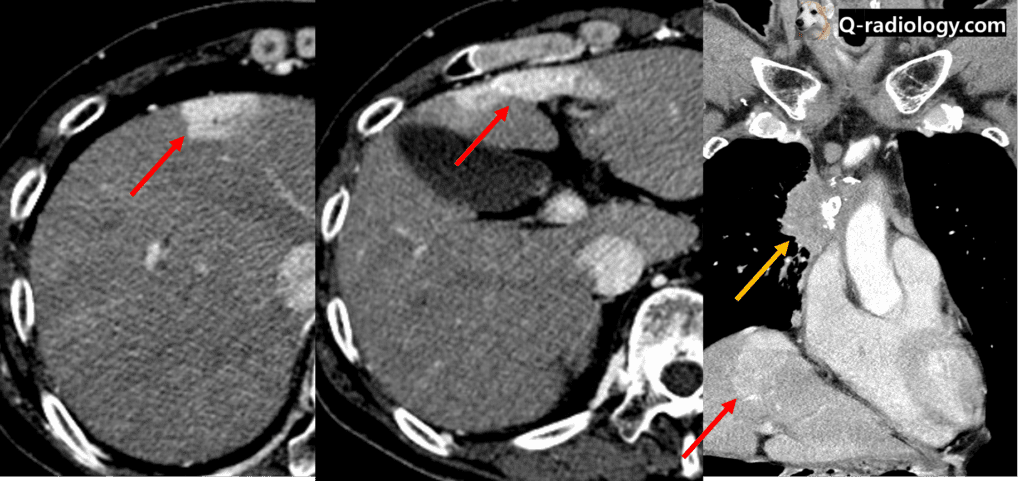
– Radionuclide uptake in liver : Hot quadrate sign (hepatic hot spot sign)
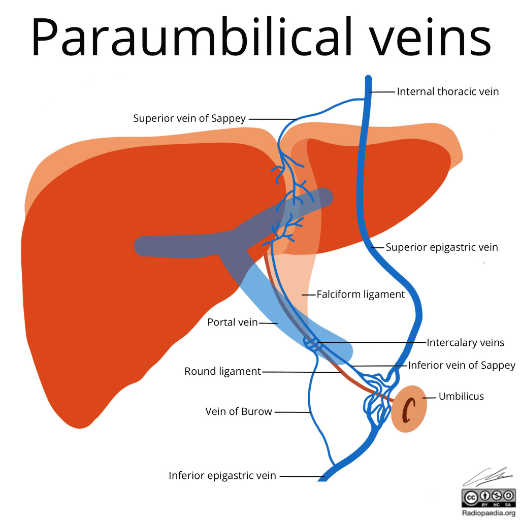
– Contrast flow
Upper limb vein ➜ collaterals (internal thoracic, superior epigastric, inferior epigastric veins) ➜ paraumbilical veins (superior/inferior veins of Sappey)
➜ 1) left lobe of the liver (direct hepatic parenchymal perfusion)
➜ 2) drainage into branches of portal vein ➜ left lobe of the liver
; Dual drainage = two different densities observed within the area of avid enhancement
PATHOLOGY
– Malignant etiologies (80-90%) : Lung cancer, metastasis, lymphadenopathy, lymphoma
– Benign etiologies (10-20%) : Granulomatous disease, iatrogenic, previous radiation therapy