Posterior Reversible Encephalopathy Syndrome
Posterior reversible encephalopathy syndrome (PRES) is a neurological condition that can cause a variety of symptoms, including headache, altered mental status, seizures, and visual disturbances.
It is characterized by reversible changes in brain function that typically affect the posterior regions of the brain, such as the occipital lobes (which are responsible for vision).
- Many etiologies with hypertension as common component
- Preeclampsia, eclampsia
- Drug toxicity (e.g. chemotherapy)
- Uremic encephalopathies
- Clinical issues
- Headache, seizure, mental change, visual symptoms
- Caution : some patients may be normotensive or have only minimally elevated blood pressure
Imaging of PRES
- General
- Patchy parietooccipital cortical/subcortical edema in patient with severe acute/subacute HTN
- CT
- Bilateral nonconfluent hypodense foci
- ± Symmetric lesions in basal ganglia
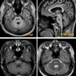
- MR
- Parietooccipital T2/FLAIR hyperintensities in 95%
- ± Basal ganglia, pontine, cerebellar involvement
- 3 patterns of hemorrhage : Focal parenchymal hemorrhage, microhemorrhages, convexity SAH
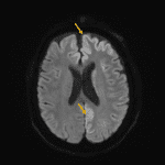
- Generally no restriction onDWI
- Variable patchy enhancement
- However, atypical imaging patterns common
Pathology
- Acute HTN damages vascular endothelium
- Failed autoregulation causes blood-brain barrier disruption
- Result = vasogenic (not cytotoxic) edema
DDx.
- Acute cerebral ischemia-infarction
- Status epilepticus
- Hypoglycemia
- Thrombotic microangiopathies (DIC, TTP, mHTN)
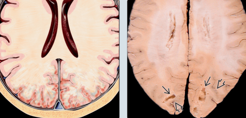
Right) Gross pathology of a patient with complicated PRES ; diffuse cerebral edema with swollen gyri
Arrow ; focal encephalomalacia secondary to infarction
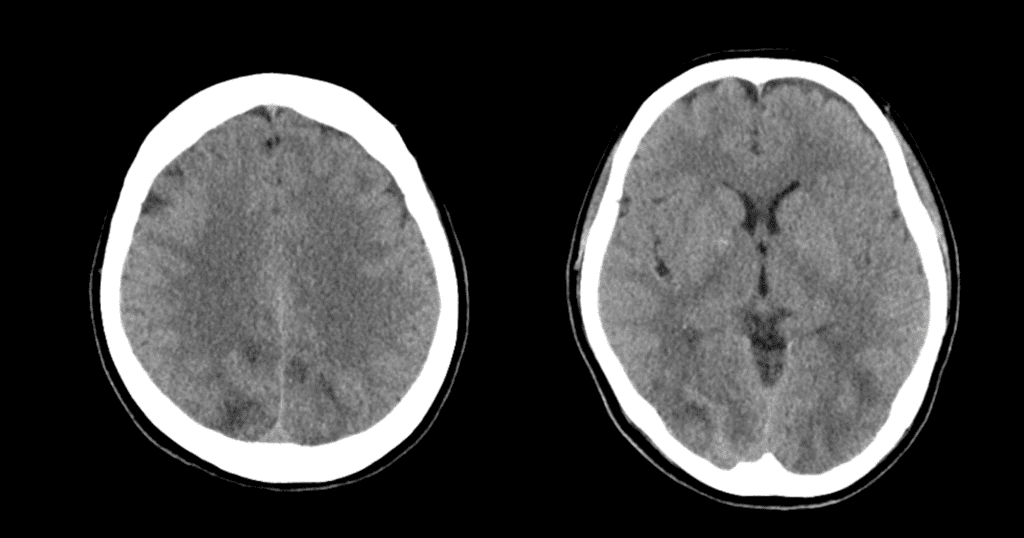
CT scan shows asymmetric subcortical edema in the occipitotemporal lobes.
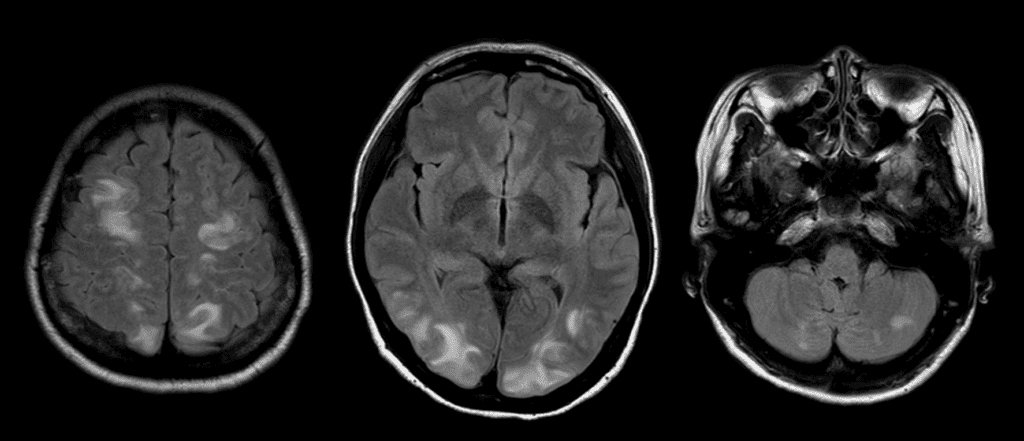
FLAIR image of the patient shows multifocal subcortical WM high signal intensities at bilateral cerebrum and cerebellum suggesting PRES.
See more about neuroimaging
Follow my instagram

Reference)
Osborn, Diagnostic imaging : brain 3rd edition, Elsevier, 911-912.