Patient history
– An 18 year-old female came to hospital due to amenorrhea with lower abdominal pain.
the patient had menarche at 13 year-old, but since 14 year-old she had no menstruation.
Mullerian duct anomaly
– From arrest of normal progression during various development of female genital tract
– 1-5% of prevalance
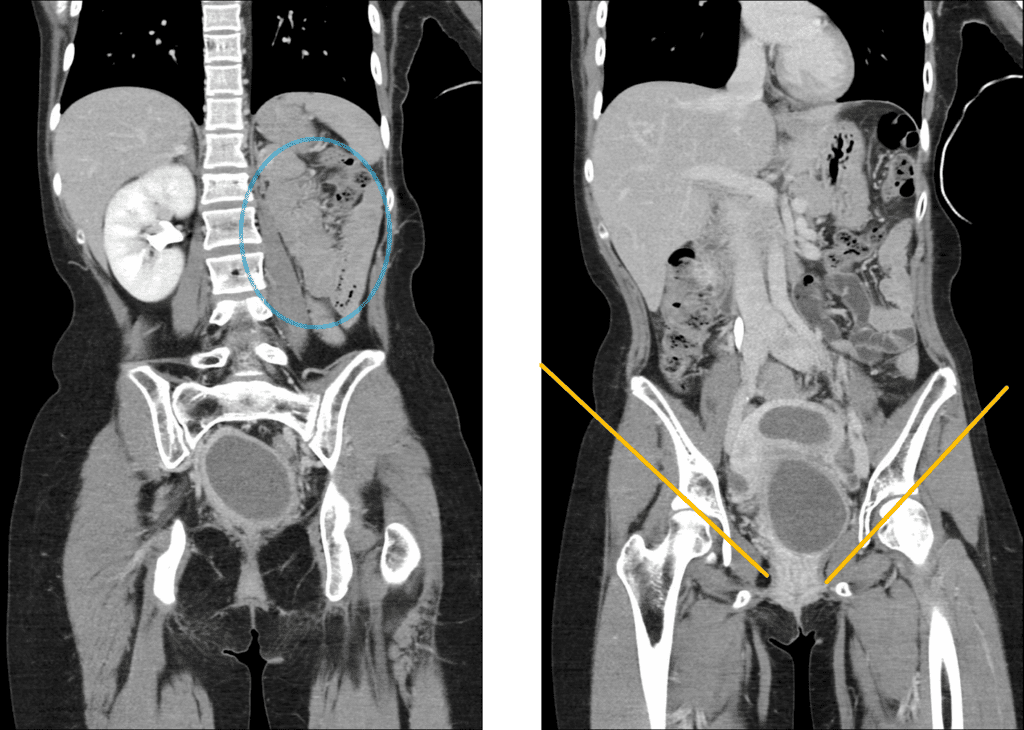
– Commonly asscociated with renal anomaly
(renal agenesis, ectopia, hypoplasia …)
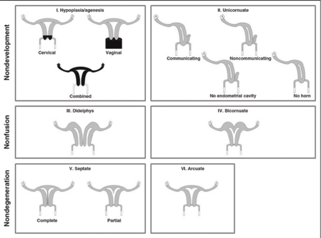
Ductal development – Nondevelopment
Ductal fusion – Nonfusion
Septal degeneration – Nondegeneration
Ductal development – Nondevelopment
Ductal fusion – Nonfusion
Septal degeneration – Nondegeneration
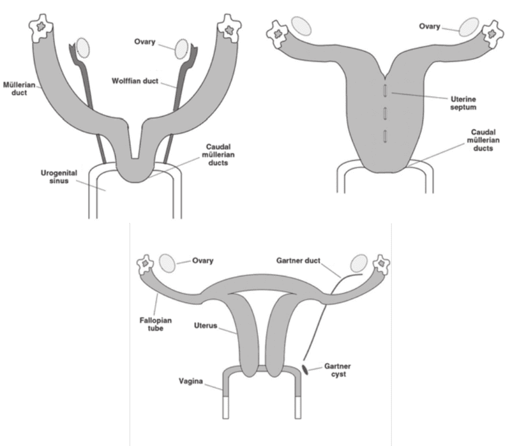
Female genital tract development
1) Ductal development
– Undifferentiated gonads with idential paired genital ducts (Wolffian & Mullerian duct)
– Absence of Y chromosome → Differentiation into ovary
– Estrogenic effect : Mullerian duct development & Wolffian duct degeneration
1-1) Nondevelopment
– Unicornuate uterus
– Rudimentary horn
– Hypoplasia/agenesis of uterus, cervix, or vagina
2) Ductal fusion
– Caudal mullerian duct fusion with intervening septum
→ Forming corpus of uterus, cervix, and upper vagina
– Upper mullerian duct form fallopian tube
2-1) Nonfusion
– Uterine didelphys
– Bicornuate uterus
3) Septal degeneration
– Degeneration of fused margin of mullerian duct
– Canalization of vaginal plate
3-1) Nondegeneration
– Uterine/vaginal septa
Longitudinal / Transverse septa
– Arcuate uterus
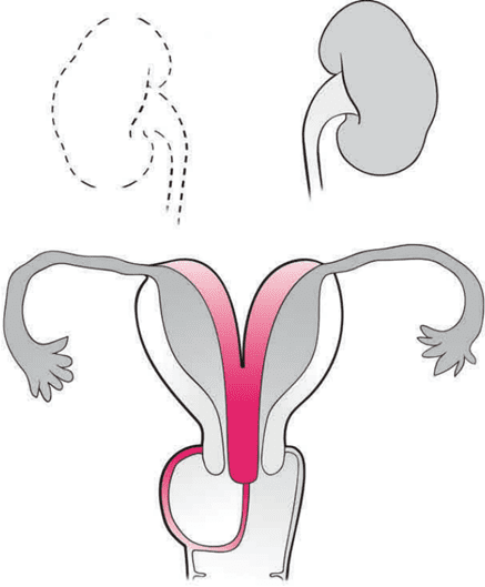
4) Herlyn-Werner-Wunderlich syndrome
OHVIRA syndrome
(= Obstructed hemivagina – ipsilateral renal agenesis)
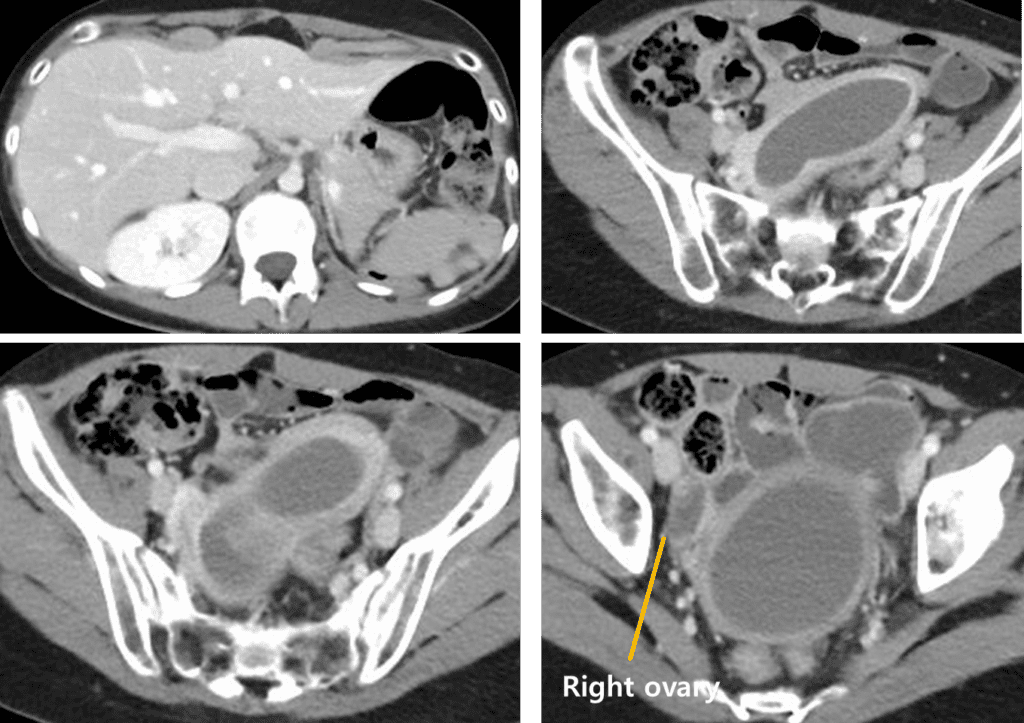
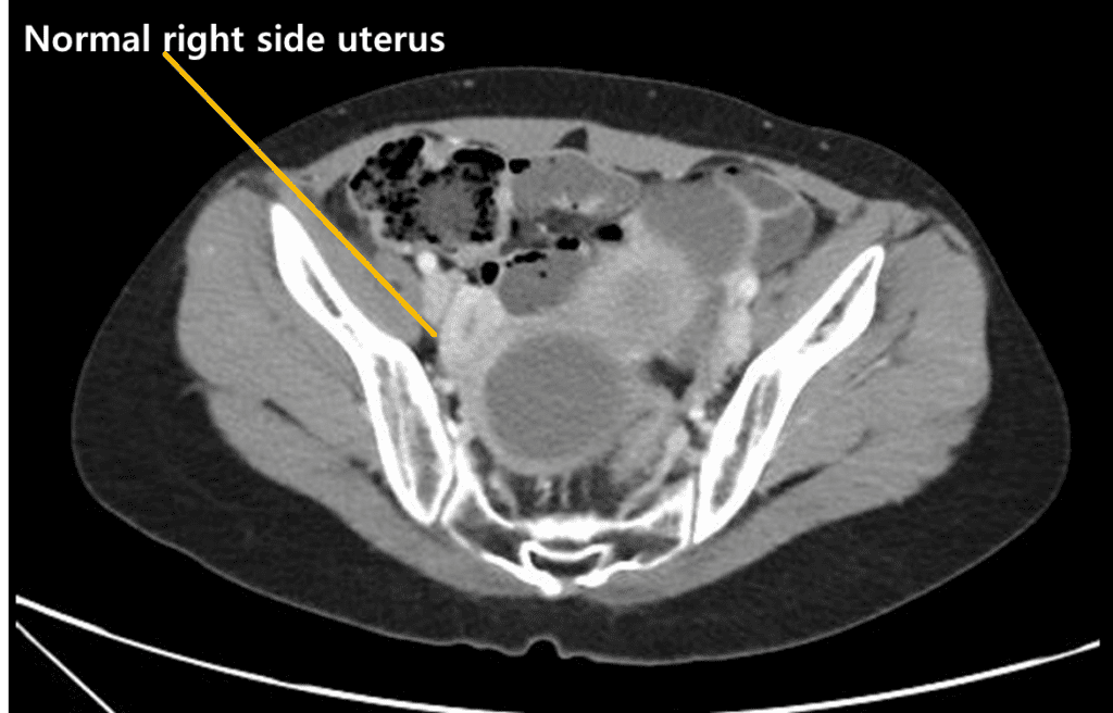
Uterine didelphys → Complete failure of mullerian duct fusion
+ Unilateral hemivaginal septum → obsturction with hematocolpus
+ Ipsilateral renal agenesis
Clinical manifestation of OHVIRA syndrome
Cyclic pelvic pain with regular menses from unobstructed side
Increased prevalence of endometriosis, pelvic adhesion
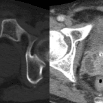
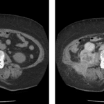
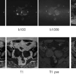
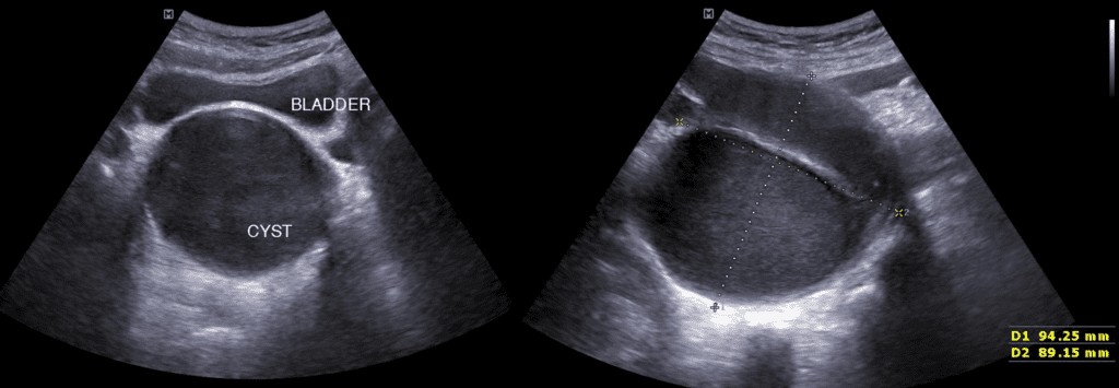
Imaging feature of OHVIRA syndrome
US: Uterine fundal cleft > 1cm (100% sensitivity & specificity)
MR :
1. The presence of unilateral obstructed hemivaginal septum
2. Unilateral obstructing distended hemivaginal horn
References)
RadioGraphics 2012; 32:E233–E250
RadioGraphics 2009; 29:1085–1103
AJR 2012; 198:302–310
Pediatr Radiol (2007) 37:657–665