Lung disease classification by nodular distribution pattern
- Small nodules with centrilobular distribution
- Infectious bronchiolitis
- Haemophilus influenzae pneumonia
- Mycoplasma pneumoniae
- Bronchogenic dissemination of pulmonary tuberculosis
- Random and patchy distribution
- Nodular bronchiectatic form of NTM pulmonary disease
- RML and LUL lingula
- Diffuse panbronchiolitis (DPB)
- Middle and lower lung zone predominance
- Symmetric subpleural distribution along with bronchiectasis
- Severe pansinusitis
- Patients from Asia (especially Korea and Japan)
; HLA-associated major susceptibility gene - Association : Sinusitis
- Subacute hypersensitivity pneumonitis
- Random and patchy distribution
- Follicular bronchilolitis
- Lymphoid follicles containing reactive germinal centers in the walls of bronchioles and occasionally pleura, especially in patients with rheumatoid arthritis (RA) and Sjogren’s syndrome
- bilateral centrilobular small nodules and peribronchiolar nodules
(mostly, <3mm) - Patchy GGO areas, bronchial wall thickening, patchy areas of low attenuation
- Random and patchy distribution
- Bronchus-associated lymphoid tissue lymphoma
- Random and patchy distribution
- Cystic fibrosis
- Infectious bronchiolitis
Caution : vascular tree-in-bud sign may be seen in …
pulmonary tumor embolisum
localized lymphangitic carcinomatosis
foreign-body-induced necrtizing vasculitis
necrotizing pulmonary vasculitis in SLE
localized pulmonary lymphatic metastasis
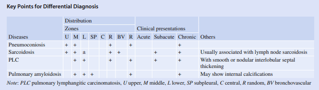
- Small nodules with perilymphatic distribution
; The presence of thickening or nodularity of the bronchovascular bundles- Pneumoconiosis
- Permanent alteration of the lung structure due to the inhalation of mineral dust and the tissue reactions of the lung to its presence
- Multiple small nodules of 2-5mm in diameter
- The nodules tend to involve mainly dorsal regions of the upper lobes and are most numerous in the right upper lobe
- When nodules coalesce to form opacities larger than 10 mm in diameter, they are called progressive massive fibrosis (PMF)
→ PMF tends to develop in the periphery of the upper and middle lung zones and often appears to migrate gradually toward the hila
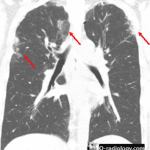
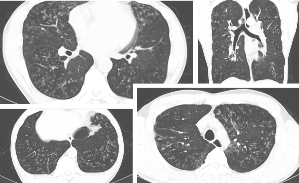
- Pulmonary sarcoidosis
- Non-necrotizing granulomatous inflammation distributed primarily along the bronchovascular bundle and lymphatics
- The granulomas predominantly involve airway submucosa and the pulmonary interstitium rather than airspaces.
- Small nodules with perilymphatic distribution (2-4mm)
- Round shape, biateral/symmetric distribution
- Subpleural and peribronchovascular interstitium and less often in the interlobular septa.
- Sarcoid nodules may coalesce over time, forming large nodules.
- Reticulonodular pattern, parenchymal consolidation, ground- glass opacity, and fibrocystic changes
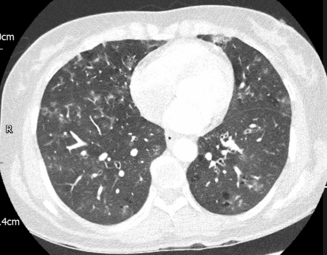
- Lymphangitic carcinomatosis
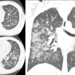
- Pulmonary alveoloseptal amyloidosis
; interlobular septal thickening, perilymphatic nodules, consolidation- Extracellular deposition of misfolded proteins in beta-pleated sheets.
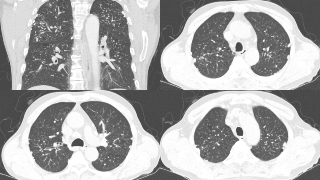
- Primary pulmonary amyloidosis is amyloid deposition in the respiratory tract without associated systemic amyloidosis
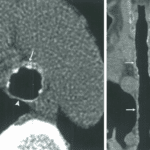
- Tracheobronchial amyloidosis
; focal or diffuse tracheobronchial wall thickening thah may be circumferential (with or without calcifications) - Nodular amyloidosis
; solitary or multiple pulmonary nodules, often with calcifications
; Amyloid nodules in the lung parenchyma are usually an incidental finding that need to be distinguished from neoplasia. They are usually peripheral and subpleural, occur more frequently in the lower lobes, may be bilateral, and range in diameter from 0.4
- Tracheobronchial amyloidosis
- Reticular opacity, interlobular septal thickening, and small nodules (2-4mm)
- Pneumoconiosis
- Small nodules with Random distribution (Miliary distribution)
; the small nodules are diffusely seen in all lobes of both lungs and affect the lungs either equally or symmetrically.- Miliary tuberculosis
- Miliary metastasis
- Disseminated fungal pneumonia (candida albicans and cryptococcosis)
- Disseminated viral pneumonia
- Sarcoidosis (miliary form)
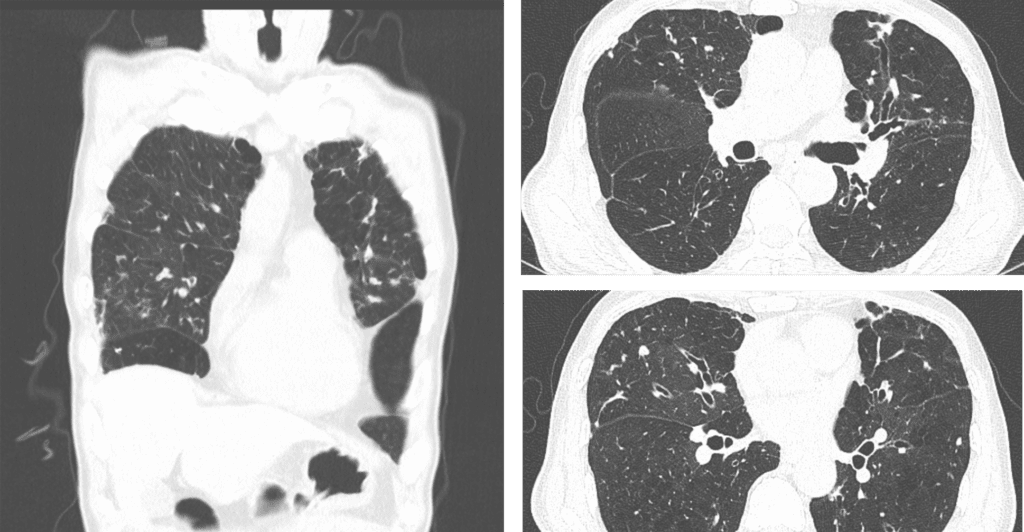
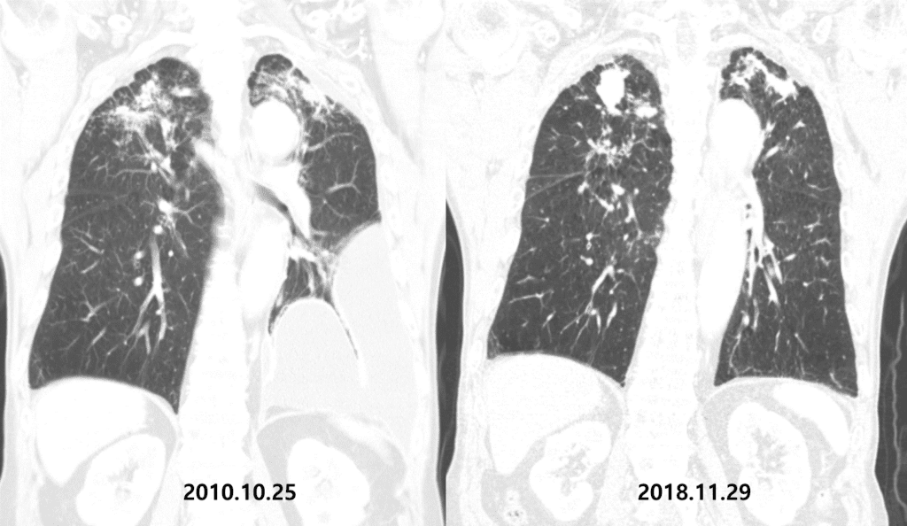
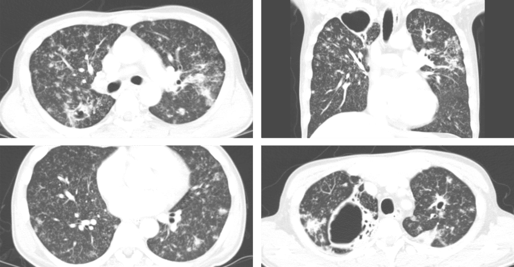
bronchial wall thickening are also seen. follicular bronchiolitis.
case from radiopaedia
Nodules slowly aggregates forming mass, mimicking malignant mass
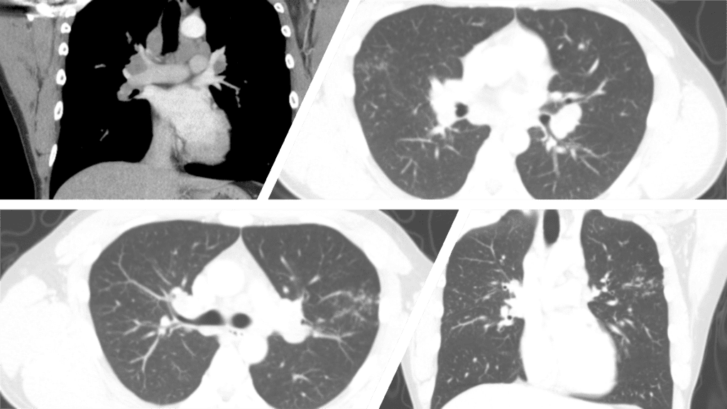
Multiple ill defined clustered nodules with GGO in LUL and RML (galaxy sign).
Bilaterally increased mediastinal and hilar lymph nodes are found.
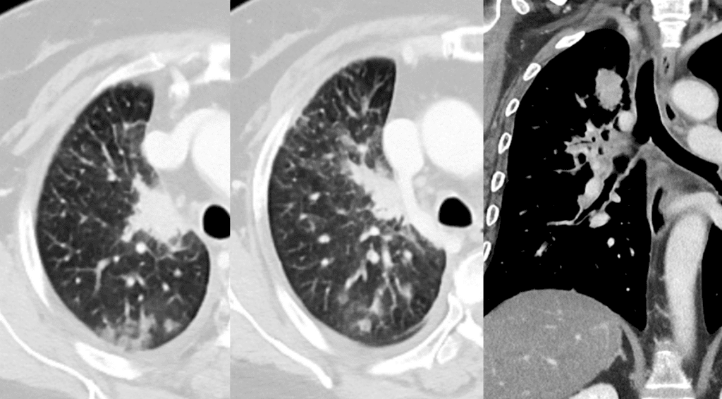
GGA, reticulation, calcified small nodules, thickening of ILS and traction bronchiectasis in both lung.
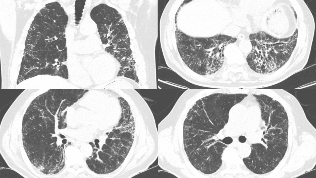
diffuse thickening and calcification from trachea to peripheral bronchi in both lung
nodules, cavitary nodules, centrilobular nodules and miliary nodules in both lung
See more about chest imaging
Follow my instagram