What is double aortic arch?
Double aortic arch is a common type of complete vascular ring, a congenital anomaly of the aorta and its branches.
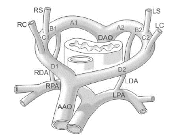
In double aortic arch, the aortic arch fails to appropriately remodel during embryology, resulting in an ascending aorta that divides anterior to the trachea and esophagus, with one arch coursing to the left, and the other coursing to the right of those structures.
The arches completely encircle the trachea and esophagus and rejoin posteriorly to form the descending thoracic aorta.
As a double aortic arch surrounds the trachea and esophagus, it exerts a compressive effect.
Pathology of double aortic arch
Frequently associated with tracheobronchial tree (trachomalacia) intrinsic abnormalities
Embryology of double aortic arch
Persistent embryological right and left 4th aortic arches
Pathophysiology of double aortic arch
Severe airway and esophageal compression
Clinical issues of double aortic arch
Severe stridor, worsening with feeding
most common symptomatic vascular ring
Treatment of double aortic arch
Thoracotomy with division of the smaller of the 2 arches
Prognosis
11% of patients required a second operation

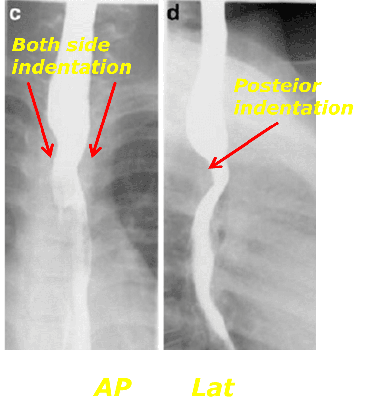
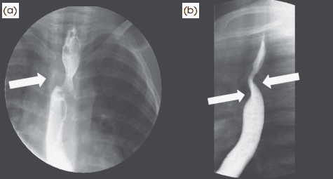
Bilateral impression of the esophagus in the reverse “S” confi guration (white arrow) is apparent on frontal view.
Anterior and posterior compression of the trachea and esophagus is seen on lateral view (white arrows).
Reference)
Charles S. White, Linda B. Haramati, Joseph Jen-Sho Chen, and Jeffrey M. Levsky (2014), Cardiac Imaging, Oxford university press
Related posts:
 Tetralogy of Fallot : Cyanotic congenital heart disease with decreased pulmonary blood flow
 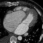 Ebstein anomaly : Cyanotic congenital heart disease with decreased pulmonary blood flow
 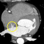 Patent foramen ovale : an Incomplete closure of the interatrial septum
 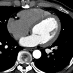 Ventricular septal defect : a hole in the interventricular septum