Restrictive cardiomyopathy
– Normal ventricular size and contractility
But a diastolic relaxation abnormality (Hallmark of restrictive CMP)
– Myocardial deposition of specific material
– Earilest form : neither dialated nor hypertrophied (seems like okay)
– Disease progressed : enlargement and a thick ventricular wall
Purpose of MR imaging of RCMP
– To determine phenotypes such as myocardial infiltrative disease
; amyloidosis, sarcoidosis, endomyocardial fibrosis, eosinophilic endocarditis
– To differentiat from constrictive perdicarditis
; RCM : thickening of myocardium including atriums (≥ 6mm)
; constrictive pericarditis : normal myocardium, thickening of pericardium (≥ 4mm), characteristic paradoxical septal motion
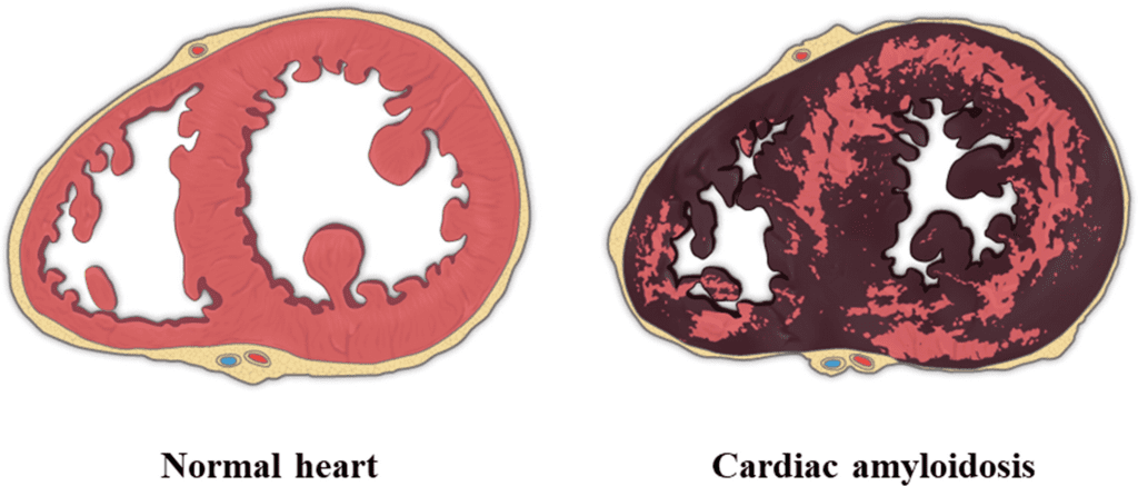
Cardiac amyloidosis
What is the cardiac amyloidosis?
Amyloidosis is the extracellular deposition of insoluble proteins and their derivatives that occurs systemically affecting one or multiple organs.
The interstitial deposition of amyloid within the heart leads to cardiac injury and is termed cardiac amyloidosis.
Cardiac amyloidosis is the most common cause of restrictive cardiomyopathy in most parts of the world.
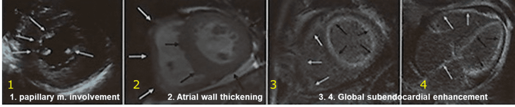
Imaging finding of cardiac amyloidosis
MR findings
– Commonly all chamber involved (especially, pathognomonic when interarterial septum and right atrial free wall by more than 6mm)
– Papillary muscles are involved (precede wall involvment)
– Thickening of the ventricular and atrial walls as well as the interatrial septum
– Global subendocardial enhancement (69%, highly sensitive, poor prognosis)
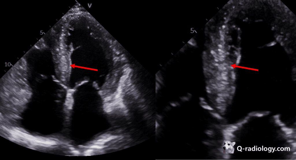
Echocardiography
– Speckled appearance of the myocardium due to amyloid deposition
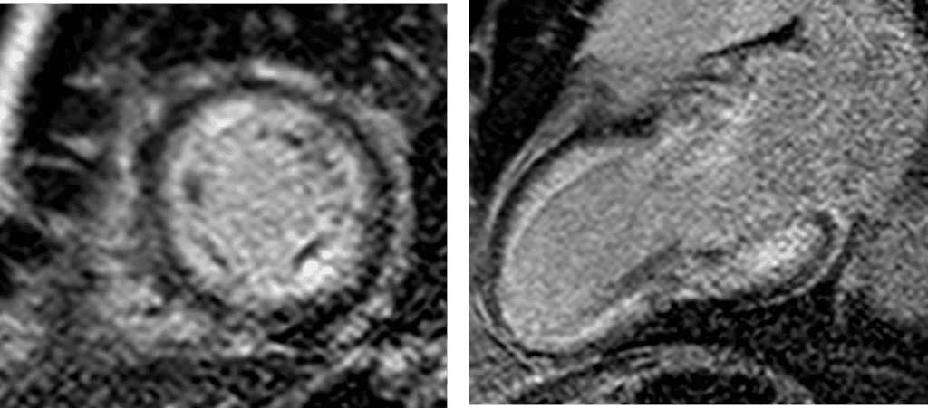
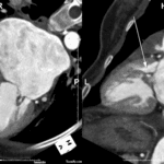
Cardiac amyloidosis, global subendocardial enhancement on DE-MRI
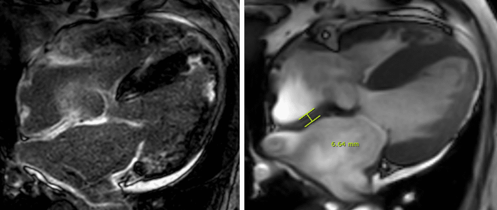
Left) global subendocardial enhancement on DE-MRI 4ch view
Right) interatrial septal thickening more than 6mm.
Reference)
Charles S. White, Linda B. Haramati, Joseph Jen-Sho Chen, and Jeffrey M. Levsky (2014), Cardiac Imaging, Oxford university press
RadioGraphicsVol. 40, No. 4 https://doi.org/10.1148/rg.2020190069