What is the papillary muscles in heart?
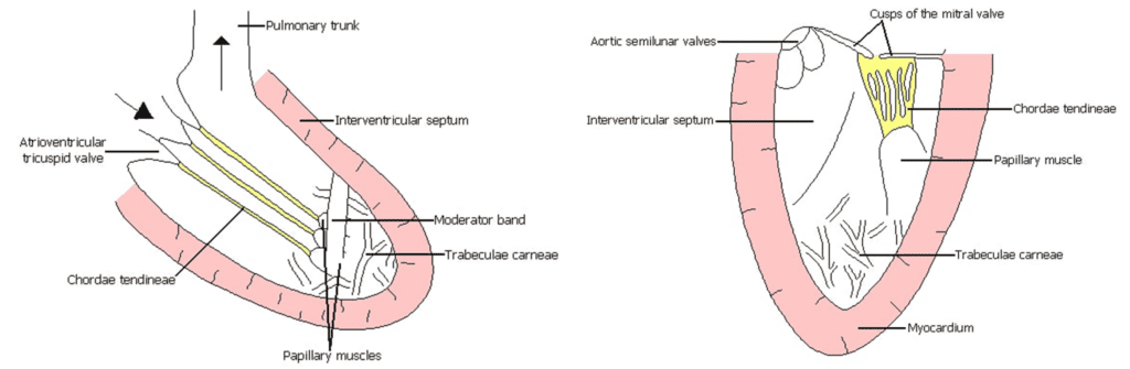
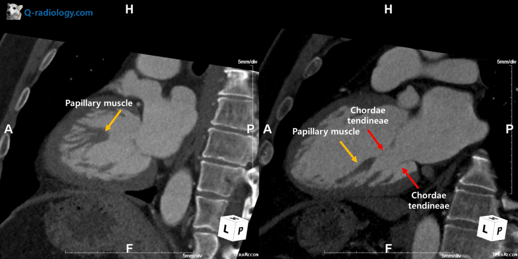
Papillary muscles are muscular structures located within the ventricular cavities, which are usually larger on the left side.
There are three papillary muscles in the RV, the anterior, posterior, and septal, and two papillary muscles in the LV, the anterolateral and posteromedial.
Papillary muscles originate from the inner wall of the ventricles in their middle or apical portions, have an elongated and tapered trunk, and continue as chordae tendineae, which then attach to the cusps of the corresponding atrioventricular valves.
What is the role of papillary muscles?
The papillary muscles play an important role in the function of the atrioventricular valves by limiting regurgitation through earlier systolic contraction than that of the ventricles and apposition of the valve leaflets.
– Early systolic contraction of papillary muscle pulls vanes of the valves inward toward the ventricle to prevent their bulging too far backward toward the atria during ventricular contracture
What happens when papillary muscle doesn’t work properly?
Papillary muscle dysfunction or rupture leads to valvular regurgitation.
– If a chorda tendinea becomes ruptured or if one of the papillary muscles become paralyzed, the valve bulges far backward during ventricular contraction, it could make blood flow to regurgitate.
Hypertrophy and a higher number of heads manifest in hypertrophic cardiomyopathy (HCM)
Reference)
RadioGraphics 2019 39:5, 1238-1263