DEFINITION
– Benign pulmonary tumor containing multiple mesenchymal tissue elements
PATHOLOGY
– Most common benign lung tumor (75%)
– Common in 40-50s
– Varying amounts of cartilage, fat, connective tissue, smooth muscle …
IMAGING
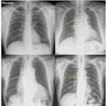
– Peripheral location (2/3)
– Endobronchial location (10%) ; obstructive symptom
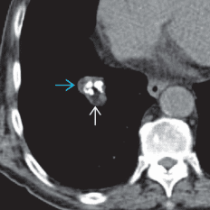
– Round or smooth lobulated mass with popcorn calcification and fat density
– Usually shows poor enhancement
– Slow growing ; less than 5mm/year
– Carney triad : a rare syndrome defined by the coexistence of three tumours
➜ multiple hamartoma / GI leiomyosarcoma / extra-adrenal paraganglioma
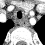
Endobronchial hamartoma
– Focal endoluminal lesion in central airway
– Internal fat &/or calcification suggest diagnosis
– Fat in endoluminal nodule only seen in airway hamartoma or lipoma
– Postobstructive findings: Atelectasis, consolidation, bronchiectasis
– Little or no uptake on FDG PET