- Slipped capital femoral epiphysis (SCFE) is defined as the displacement of the femoral head relative to the femoral neck and shaft.
- Boys> Girls (2.5:1)
- Affected children are frequently obese.
- In approximately one-third of patients, there is bilateral (but not typically simultaneous or symmetric) involvement of the hips.
- History of trauma in approximately 50% of patients.
- Imaging finding
- Radiograph
- The earliest : Widening and irregularity of the physis
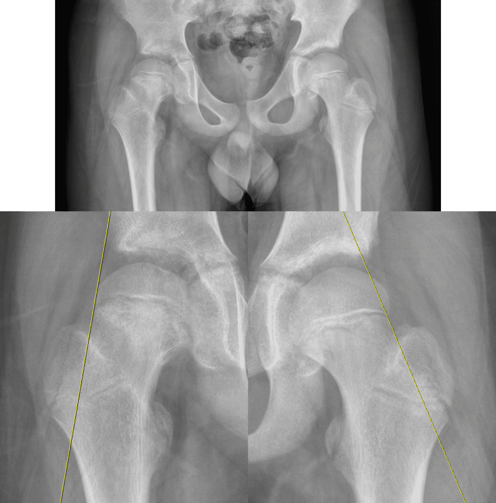
- Trethowan sign ; See below
- Chronic slip : sclerosis may be seen in the medial femoral neck with bone remodeling (buttressing).
- CT and MR may also be used to make the diagnosis of SCFE.
- Diffuse or globular physeal widening is the earliest evidence of SCFE on MRI.
- Radiograph
- Due to complete loss of osseous continuity, SCFE is associated with high incidence of avascular necrosis as a complication.
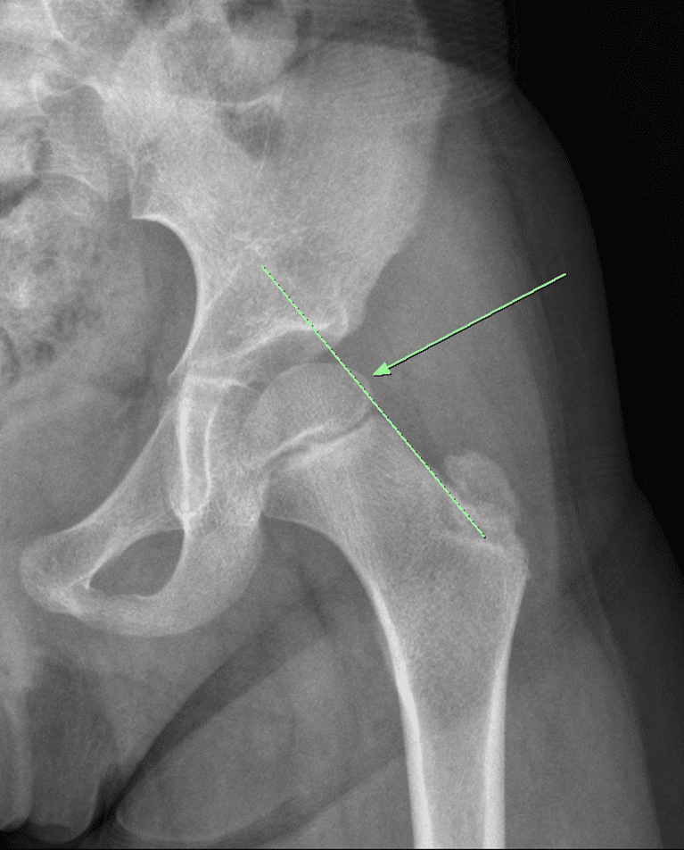
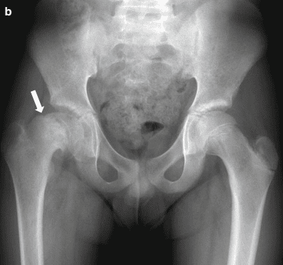
Anteroposterior radiograph in a 12-year-old boy with many months of right thigh discomfort shows slipped femoral epiphysis and ill-defined sclerosis in the right proximal metaphysis with bending of the femoral neck.
Note the bony hump ( arrow ) in the superior portion of the femoral neck, suggesting remodeling of the femoral neck.
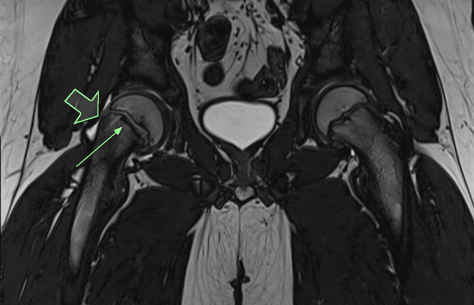
Physeal widening and epiphyseal slippage in right femoral head with surrounding bone marrow edema