What is pseudomyxoma peritonei ?
Diffuse intraperitoneal accumulation of gelatinous mucinous implants due to rupture of mucin containing neoplams (most commonly appendiceal mucinous neoplasm)
IMAGING
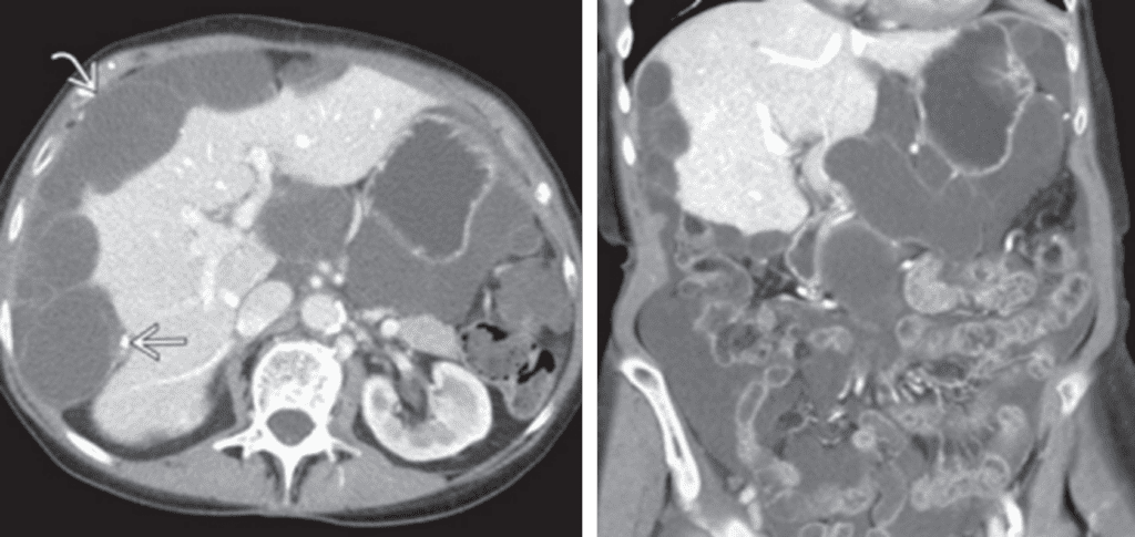
• CT: Low-attenuation masses (usually < 20 HU) scattered throughout peritoneum
– Frequently associated with loculated ascites of similar attenuation to individual implants
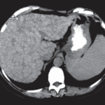
– Implants cause mass effect on liver and spleen, producing characteristic “scalloped” appearance
– Implants may demonstrate curvilinear calcification
– Dominant cystic or solid mass often present in right lower quadrant (in expected location of appendix)
– Metastases to ovary are common, so cystic masses in ovaries may not represent primary ovarian neoplasm
– Imaging findings of bowel obstruction
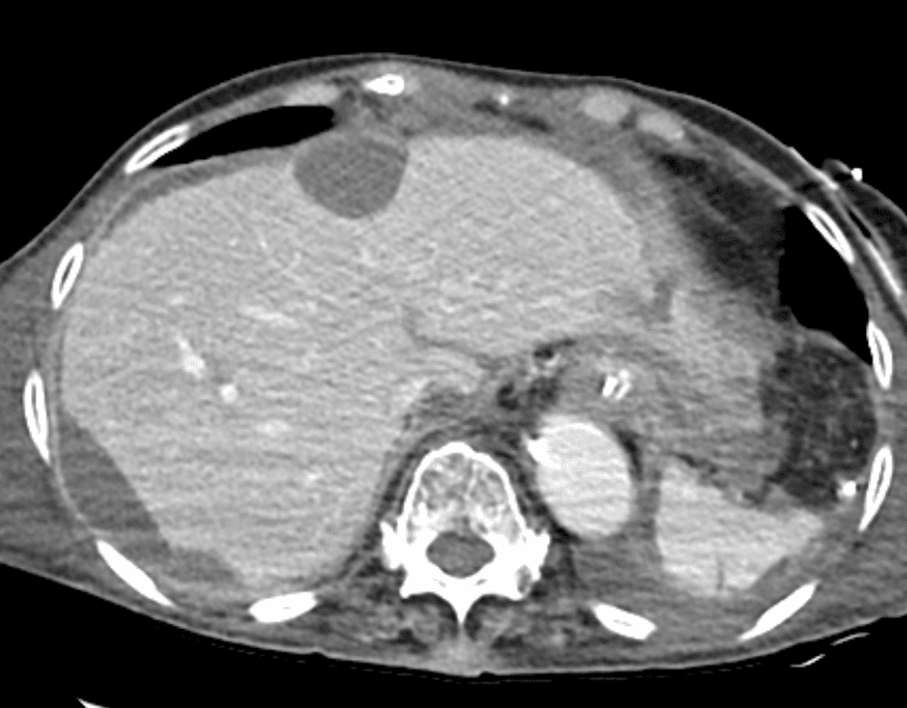
characteristic scalloped appearance of liver is shown.
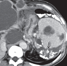
Calcifications, often curvilinear and peripheral, are not uncommonly seen with implants in pseudomyxoma