Interventional treatment for post traumatic high flow priapism
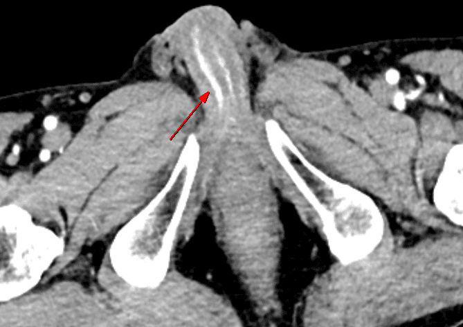
- Characteristic CT finding
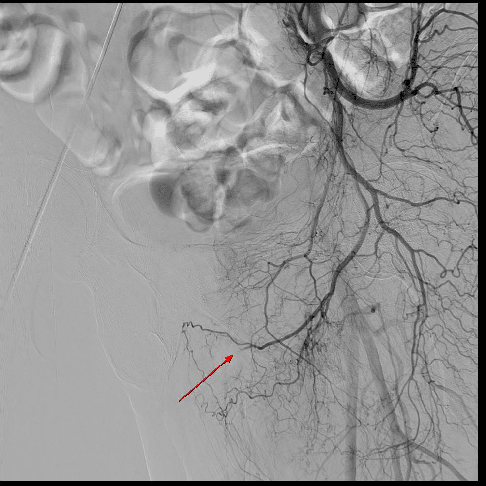
- Angiographic finding
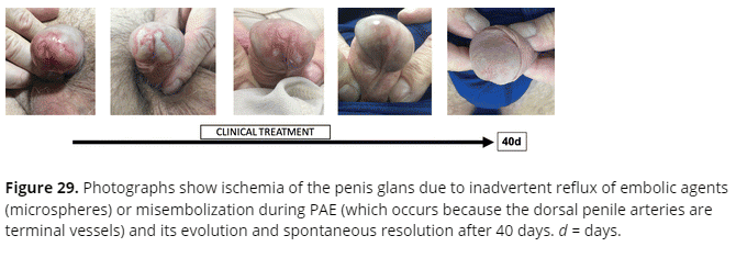
- Embolization technique preventing complication
The patient reported that he fell down, hitting his buttocks on the floor after slipping off the edge of a chair at home. Approximately two hours later, while undressing to shower, he discovered blood on his underwear and observed hematuria during urination, prompting a visit to the emergency room. The patient states that he is not experiencing any pain.
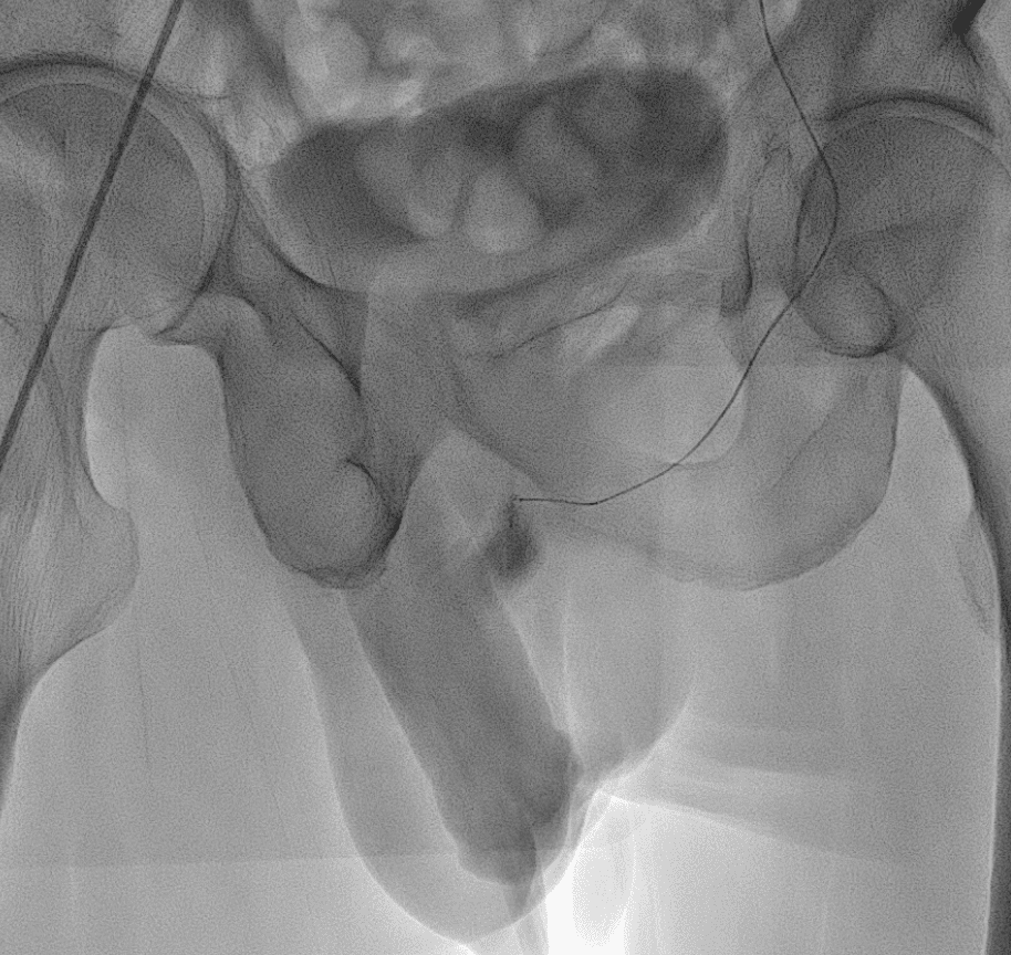
The patient’s penis was not erect, but showed signs of swelling.
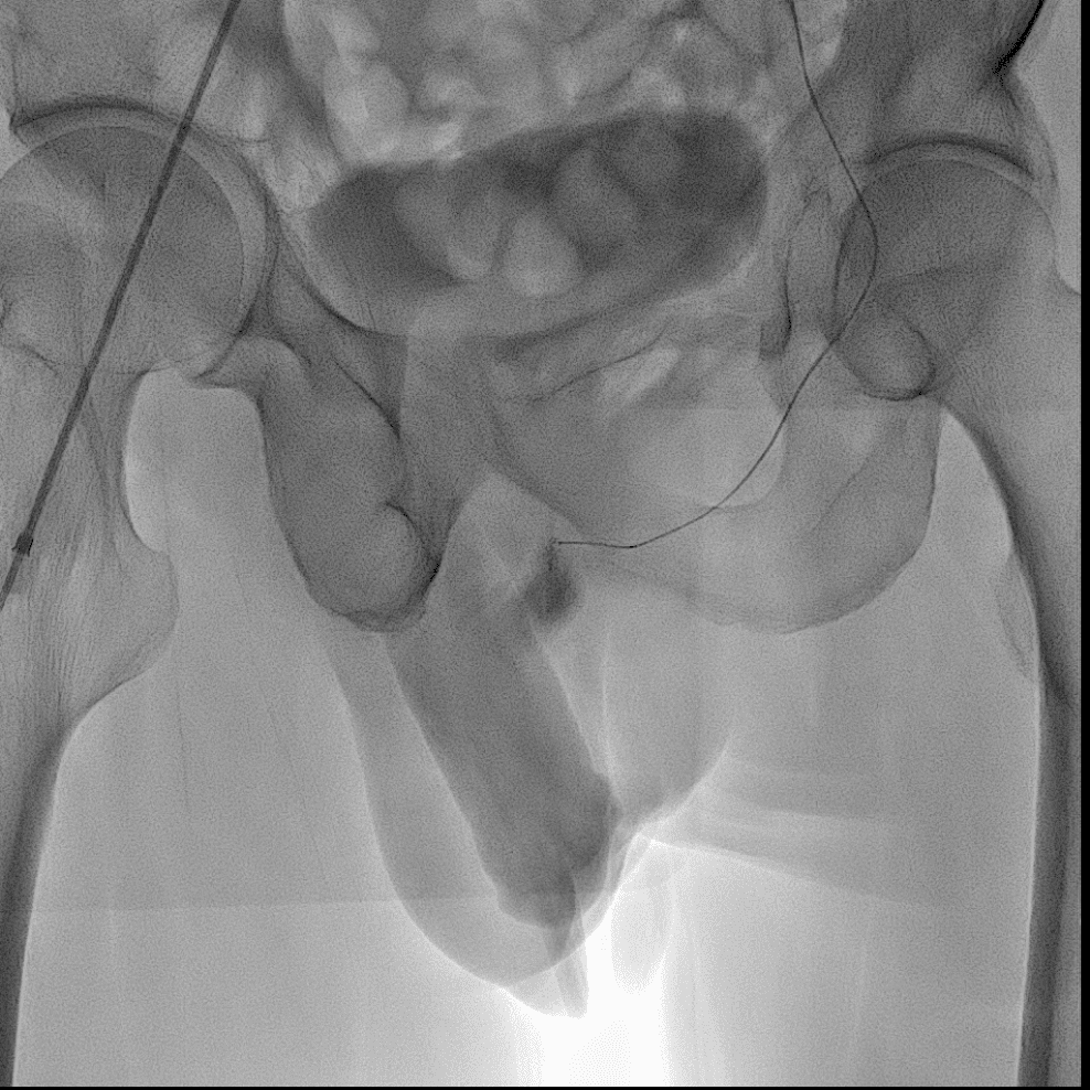
The urologist tentatively diagnosed the condition as nonischemic priapism and requested that diagnostic angiography be performed in the angioroom, followed by any necessary treatment.
The selection of the culprit artery has been completed, and treatment now needs to be initiated. However, as it involves the dorsal penile artery, there is a risk of complications such as penile necrosis if embolic material overflows during treatment and inadvertently embolizes unintended vessels, as noted in the excerpt from the journal below.

Therefore, a somewhat novel method of embolization was decided upon. The concept involves using a gelfoam particle as a plug. First, the culprit artery is superselected, and the microcatheter is flushed with saline. Next, a sufficiently large gelfoam particle is mixed with contrast agent using forceps, ensuring only one particle is used. Then, this single gelfoam particle mixed with the contrast agent is placed at the entrance of the microcatheter. After confirming the proper positioning of the gelfoam particle at the microcatheter entrance, it is pushed in using a 1cc syringe filled with contrast agent. Once the culprit artery is embolized by the gelfoam particle, even if the contrast agent overflows, the use of only one particle ensures that no other vessels are obstructed.
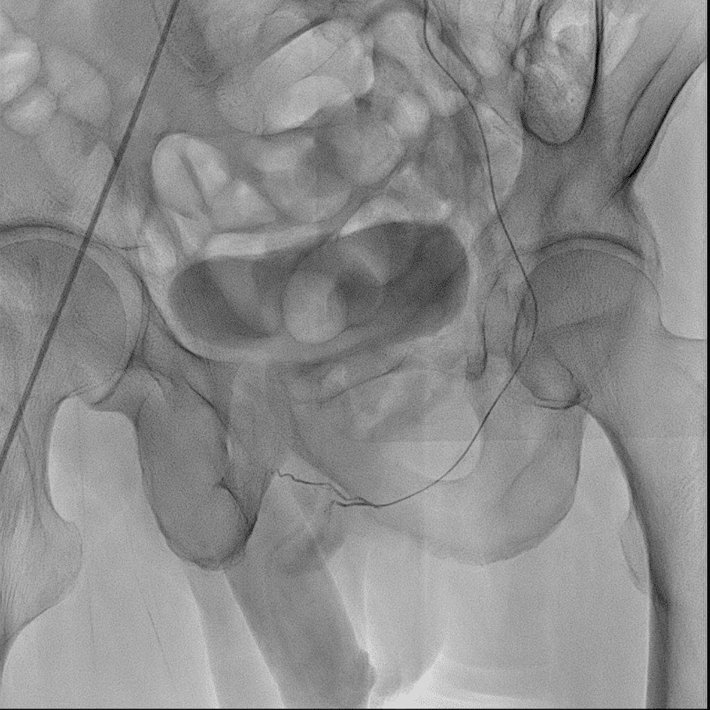
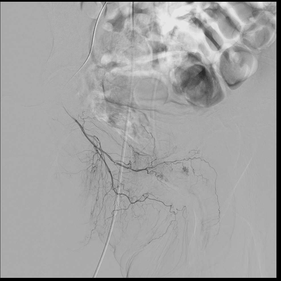

In reality, some minor hypervascularity remained, but due to concerns about potential complications, it was decided to not treat it further and to observe its progress.
priapism
Related posts:
 Today’s case 9 – IVC filter removal : how to remove filter without snaring hook
  Coil Assisted Retrograde Transvenous Obliteration (CARTO): Treatment of variceal bleeding with interventional procedure (CARTO)
  Today’s case 3 – PICC (peripherally inserted central catheter)
  Today’s case 4 – Iatrogenic bleeding of deep femoral artery while insertion of A-line