Diagnosis of mucinous carcinoma at initial biopsy is prone to sampling error, with mis-classification in approximately 25% of cases.
However, this tumor subtype has a classic appearance at MRI, allowing the radiologist to diagnose it confidently and accurately in almost all cases.
Extracellular mucin is T2 hyperintense; thus, mucinous carcinomas have significantly higher T2-weighted signal than do nonmucinous tumors.
Using T2-weighted signal, MRI is 96–97% accurate at predicting the mucinous histologic type with sensitivities of 94–100% and specificities of 95–98%.
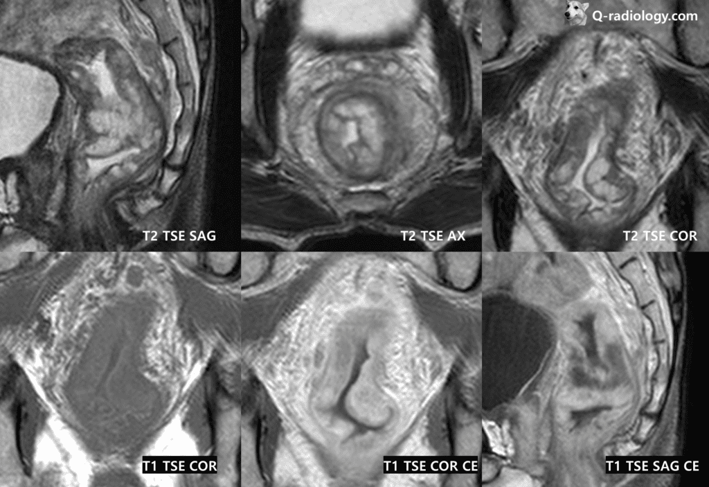
T1WI and T1CE show heterogeneously enhancing mass in rectum.
Reference)
Wnorowski, AM, Menias, CO, Pickhardt, PJ, Kim,
DH, Hara, AK and Lubner, MG. Mucin-containing rectal carcinomas: Overview of unique clinical
and imaging features. American Journal of Roentgenology. 2019; 213(1): 26–34. DOI: https://doi.
org/10.2214/AJR.18.20864