What is Metachromatic leukodystrophy?
Metachromatic leukodystrophy (MLD) is a rare genetic disorder that affects the central nervous system.
It is a type of leukodystrophy, which is a group of disorders that affect the white matter of the brain and spinal cord.
In people with MLD, lack of arylsulfatase A(ARSA) leads to a buildup of sulfatide in the brain and other parts of the body.
This can cause a wide range of symptoms, including developmental delays, behavioral changes, difficulty walking and speaking, and eventually, loss of mental and physical abilities.
MLD is a progressive disorder, meaning that it typically gets worse over time. There is currently no cure for MLD, but treatment can help manage some of the symptoms and improve quality of life.
• Lysosomal storage disorder caused by ↓ arylsulfatase A (ARSA) resulting in central (CNS) and peripheral (PNS) nervous system demyelination
• 3 clinical forms: Late infantile (most common), juvenile, adult
IMAGING
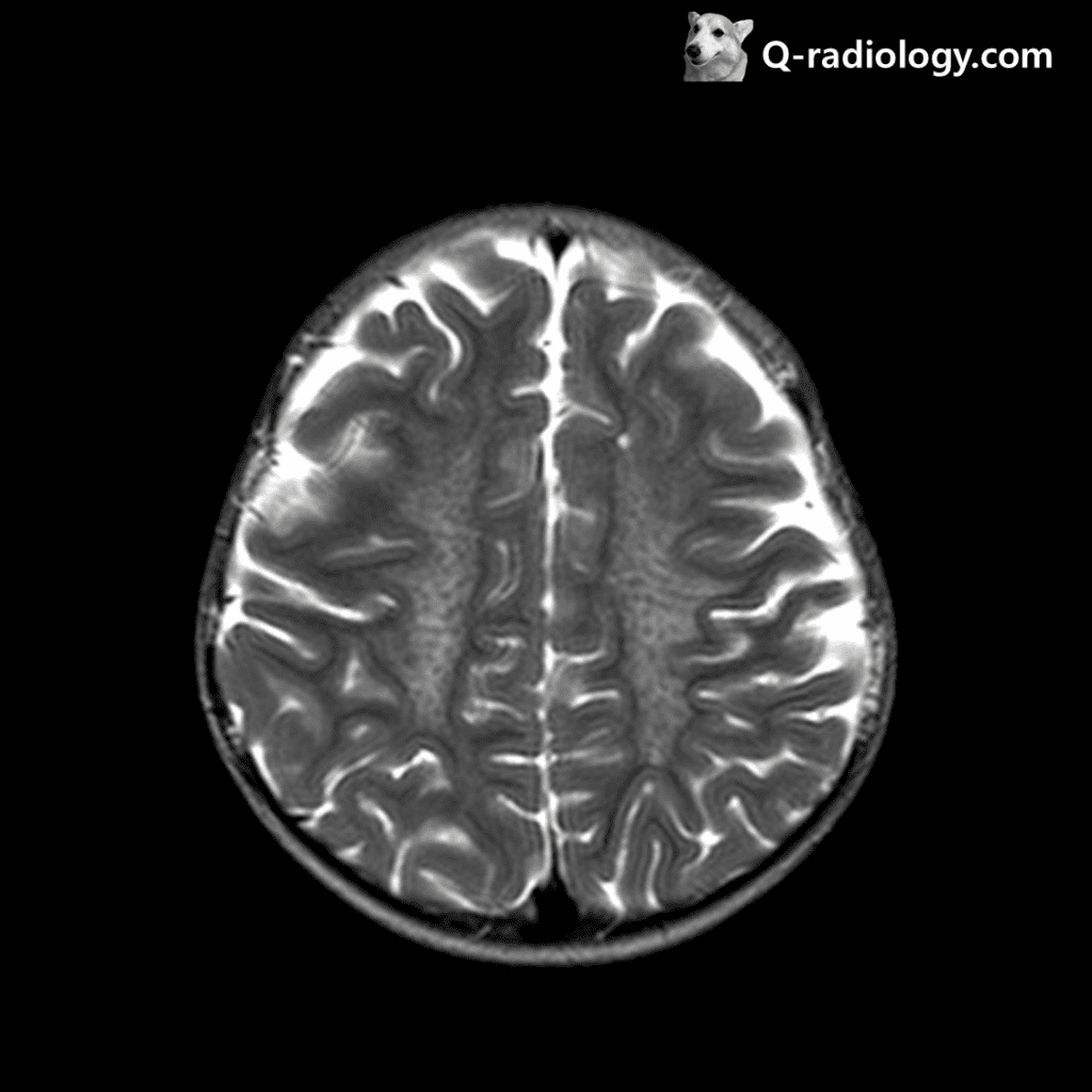
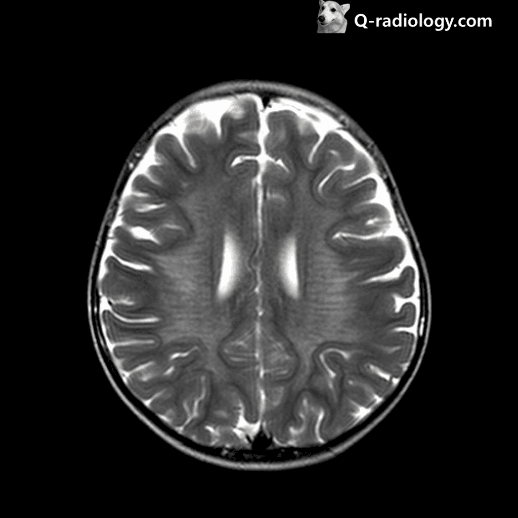
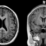
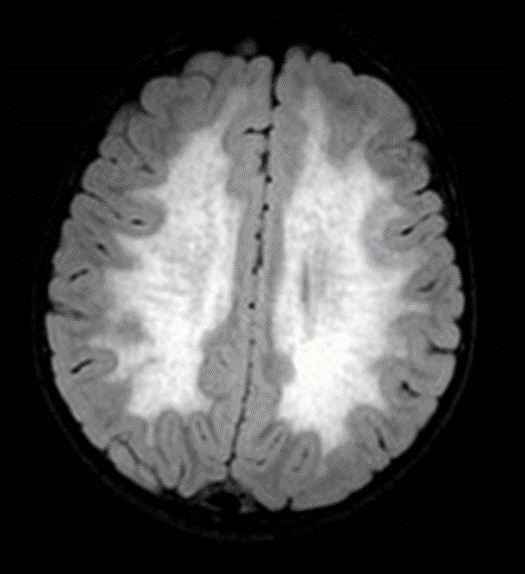
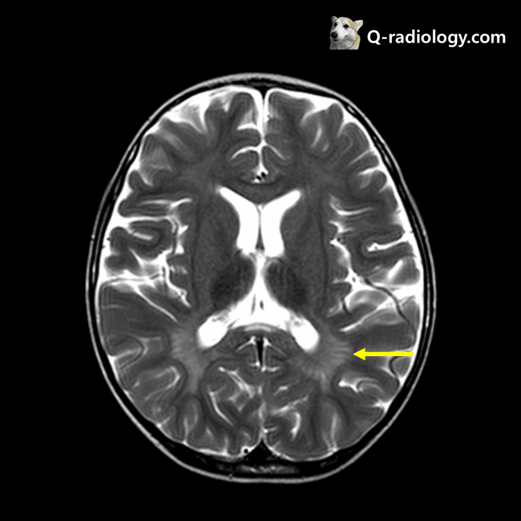
• Best diagnostic clue: Confluent butterfly-shaped ↑ T2 signal in deep cerebral hemispheric white matter
◦ Early: Spares subcortical U-fibers
◦ Late: Involves subcortical U-fibers
• Sparing of perivenular myelin = “tigroid” or “leopard” pattern
• No WM enhancement
◦ Reports cranial nerve, cauda equina enhancement
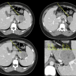
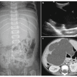
Because sulfatide accumulates also in the wall of the gallbladder, cholecystitis is a recognized complication
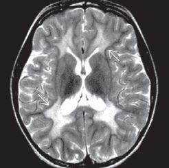
Image from Diagnostic imaging, pediatric neuroradiology, 2nd edition
The sparing of perivenular myelin is thought to account for this appearance
Metachromatic leukodystrophy