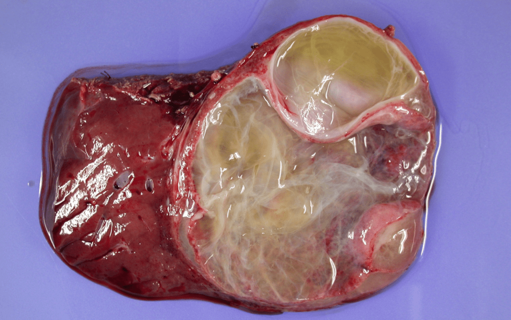
Mesenchymal hamartoma
- Mesenchymal hamartoma is the second most common benign liver mass in children after vascular tumors.
- Most are discovered before 5 years of age
- Boys > Girls
- Diagnosed on prenatal ultrasound
- Generally considered a congenital lesion related to a developmental anomaly
- Most common clinical presentation : Painless abdominal distension with normal AFP level
- The mass may be pedunculated and attached to the inferior surface of the liver
- Imaging finding
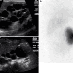
- USG
- Multicystic, heterogeneous masses with septa of variable thickness
- When the cysts are tiny – mimics a solid lesion
- Gelatinous contents or hemorrhage ; can be seen as hypoechos in the cyst
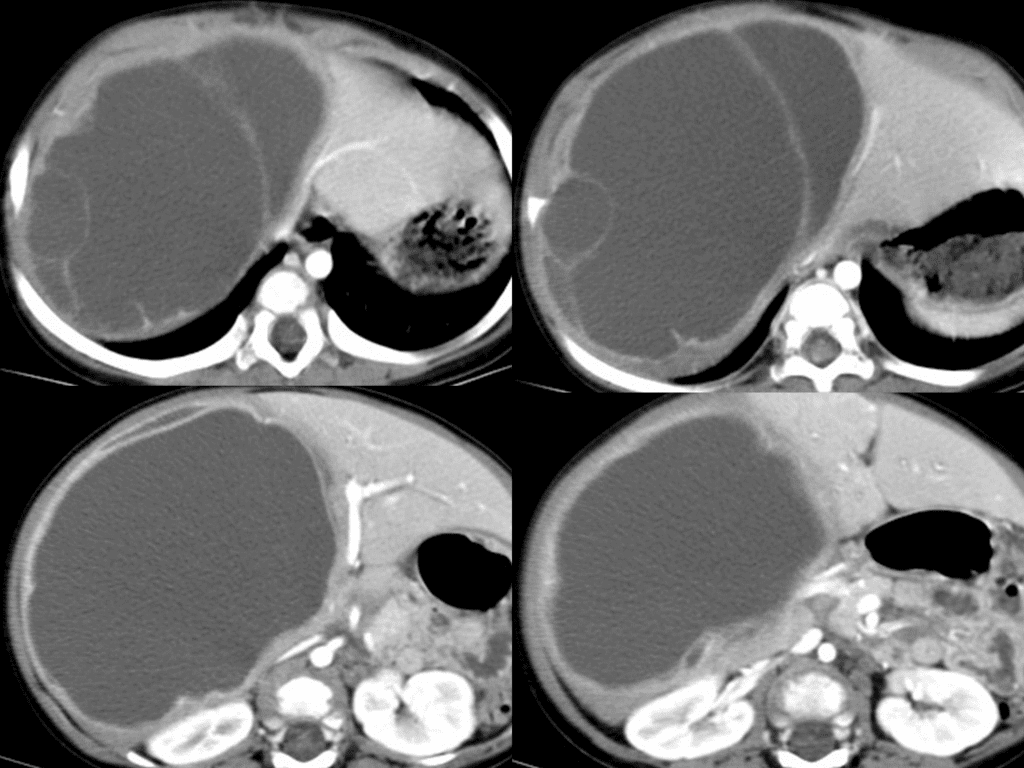
- CT
- Complex cystic masses
- Septations and solid components are enhanced
- MRI
- Cystic regions are hypointense on T1WI and hyperintense on T2WI (of coursely)
- Other portions show variable signal depending on components
- USG

Mesenchymal hamartoma in a 9-month-old boy.
( a ) Plain abdominal X-ray shows huge soft-tissue opacity in right upper abdomen.
( b ) Longitudinal US image of liver shows huge and multilobulated cystic mass originated from the liver.
( c ) Contrastenhanced abdominal CT also demonstrates multilobulated cystic mass from the liver
See more about pediatric imaging
Follow my instagram