- Second most common histologic type (10~15%)
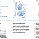
- Higher incidence of multifocality, multicentricity, and bilaterality
- E-cadherin negative
- Indian file appearance
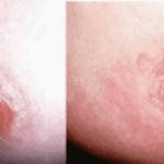
- Common cause of missed or delayed diagnosis
- Not common desmoplastic reaction ➨ US, MR is better than MG
- Larger at diagnosis than IDC
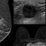
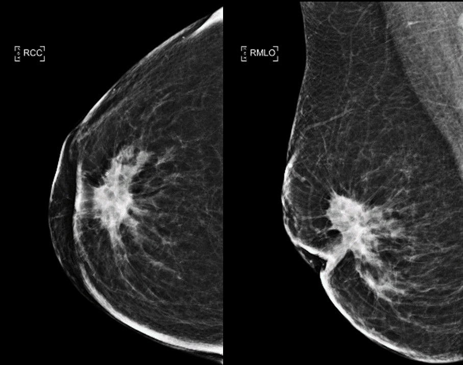
- Imaging finding
- MG : Spiculated mass (m/c), isolated architectural distortion, calcification rare
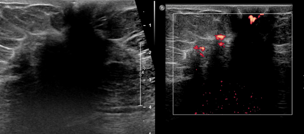
- US : Irregular hypoechoic mass, marked posterior shadowing
An 59 year old woman presented with large right breast mass
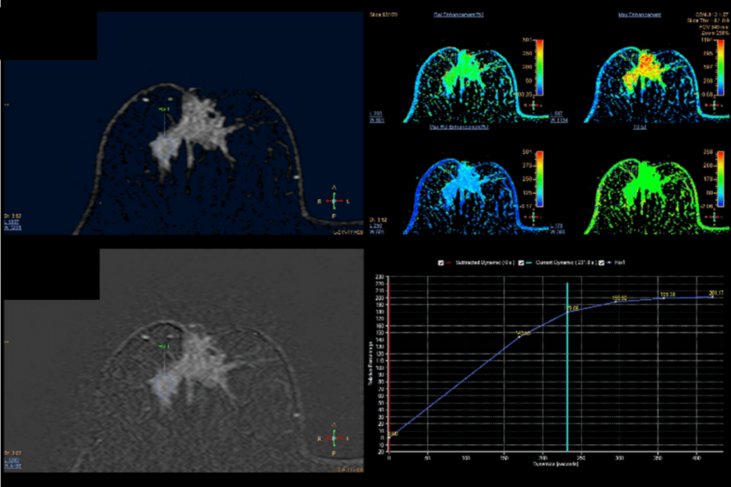
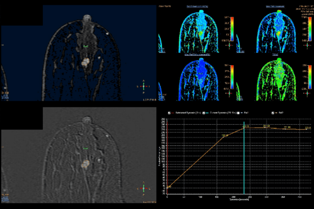
Note that invasive lobular carcinoma has tendency of multifocality, multicentricity, and bilaterality