** Neuronal migration disorder **
- Heterotopia
- Abnormal location of neurons
- Seizure disorder
- Isointense with gray matter
- No mass effect, edema, or enhancement
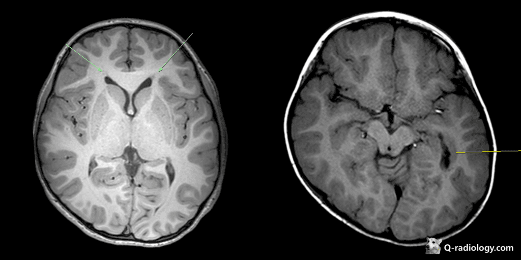
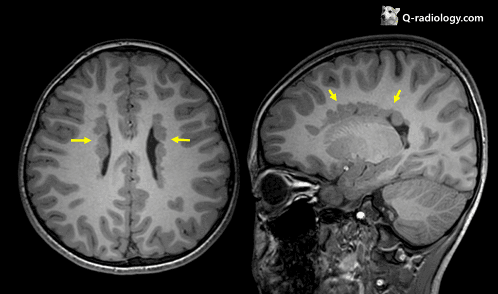
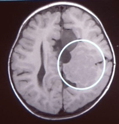
Nodular heterotopias
– subependymal heterotopia: most common
– subcortical heterotopia
Diffuse heterotopias
– band heterotopia: Now this entity is classified as a spectrum of lissencephaly
; also known as double cortex heterotopia and X-linked lissencephaly (chromosome Xq22.3)
; double cortex sign : thin interface of WM between the band heterotopia and the cortex
– lissencephaly: types 1 and 2
; Cause : arreseted neuronal migration (4 layers, thick – normally 6 layers in brain!!)
; Definition (1) Agyria : complete lissencephlay (2) Pachygyria : incomplete lissencephaly
; Spectrum : Agyria – mixed agyria/pachygyria – pachygyria – subcortical band heterotopia
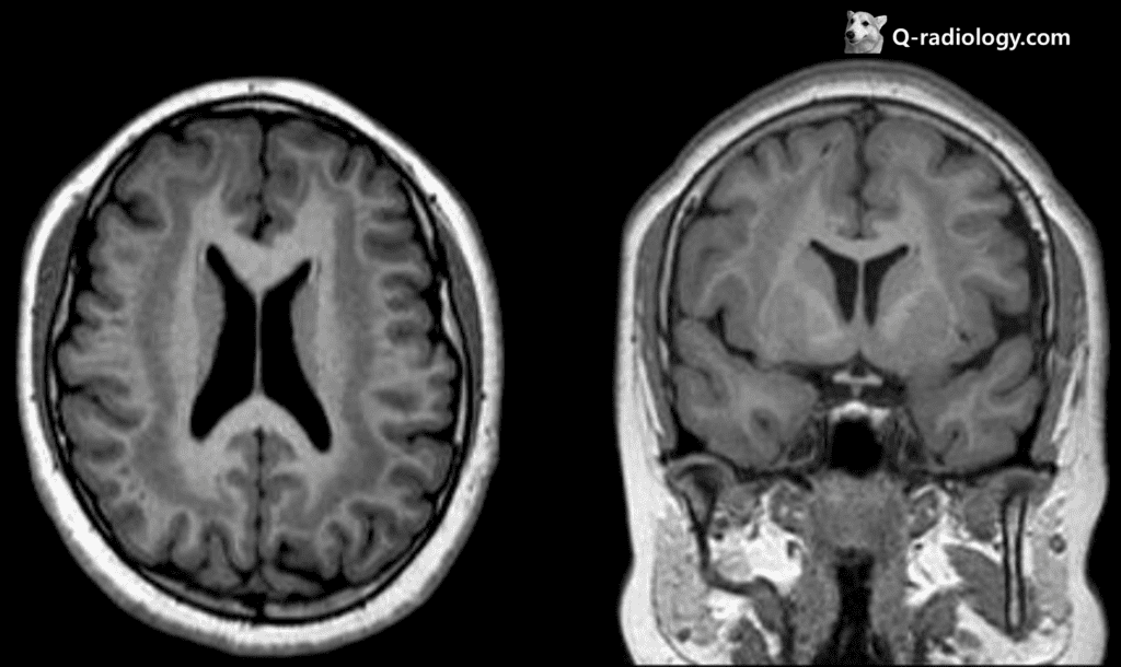
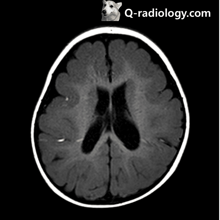
Imaging of lissencephaly
– smooth brain surface
– thick gray matter
– underdeveloped insula
– vertical orientation of sylvian fissure
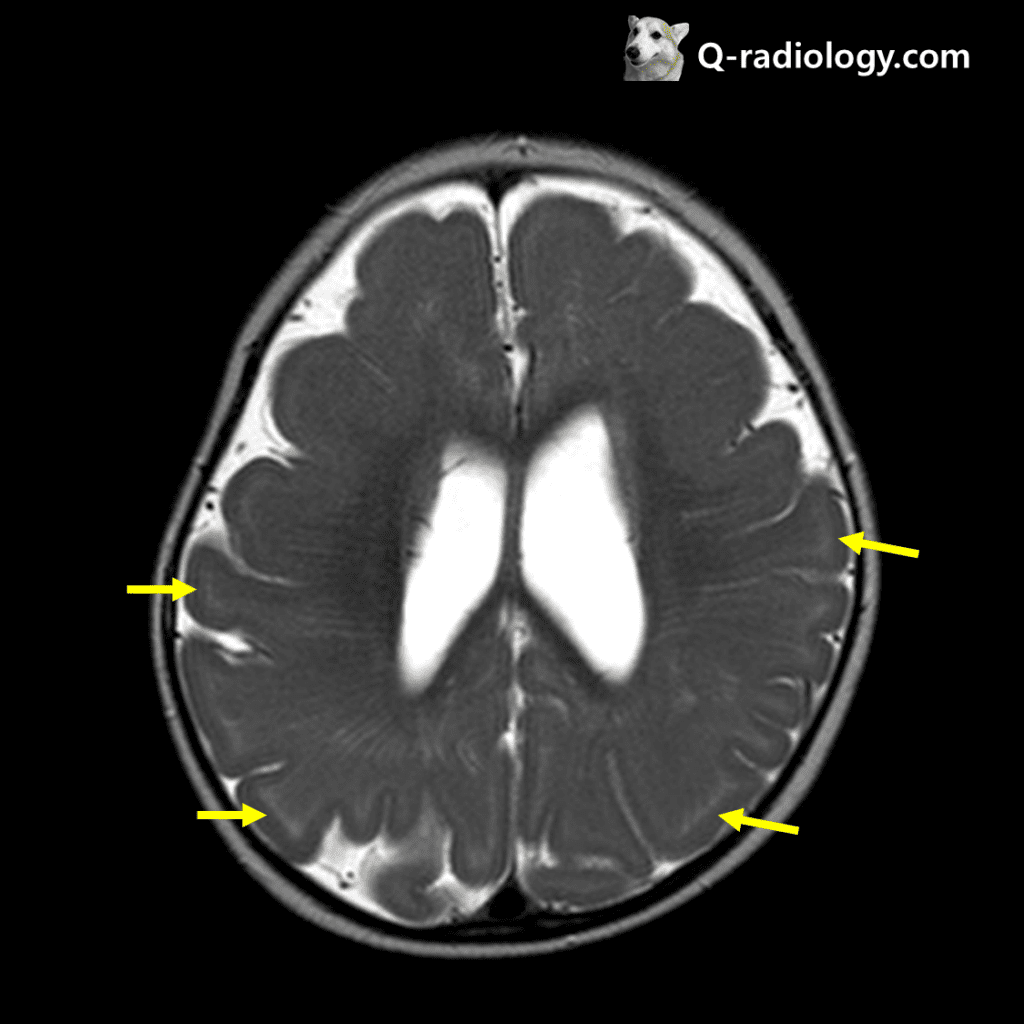
High signal intensity bands are shown, cell sparse layer (T2WI)