- 1) Fat necrosis
- Lucent mass with dystrophic calcifications
- Associated with surgery or trauma
- Follow up in 3-6 months : typically decrease in size
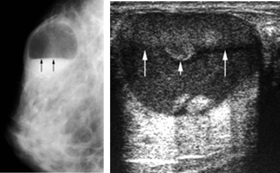
- 2) Oil cyst
- An area of focal fat necrosis becomes walled off by fibrous tissue.
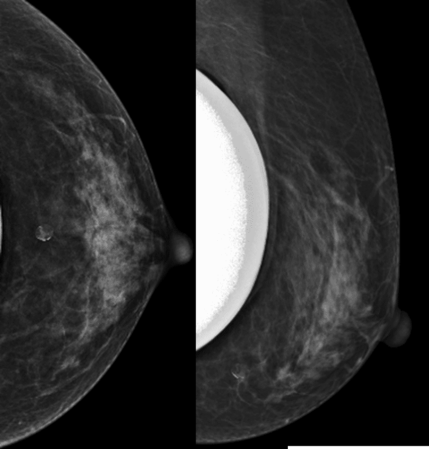
- 3) Galactocele : Fat fluid level on 90′ lateral view
- M/C benign breast lesions in lactating women
- Cystic lesion lined by cuboidal epithelium that contains milk
- More frequently occur after cessation of breast feeding
- Asymptomatic, palpable lump
- Galactoceles may be not a/w pregnancy or lactation
- Imaging finding
- Mammography
- depends on the density and viscosity of the fluid (amount of fat, proteinaceous material)
- Pseudolipoma, Pseudohamartoma
- Cystic Mass with Fat-Fluid Level : ML view
- US
- Cyst, complex cystic, mixed, solid Fat-fluid level, fluid-fluid level
- Mammography
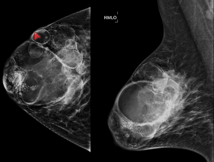
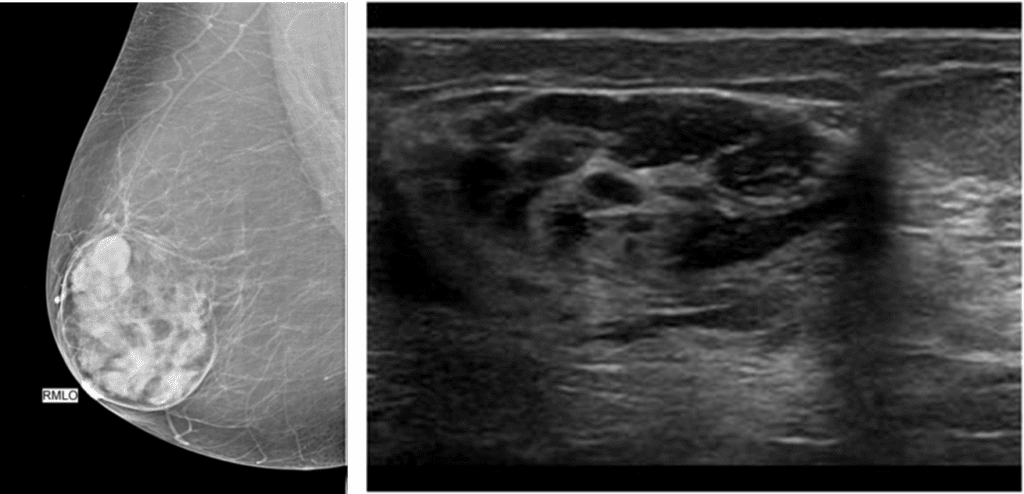
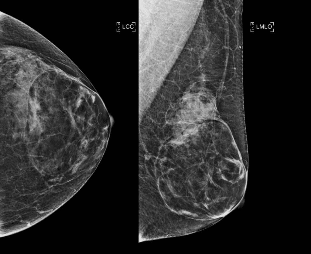
- 4) Hamartoma
- Localized overgrowth of fibrous, epithelial, and fatty elements
- “breast within a breast”
- BI-RADS C2
- Adenolipoma, fibroadenolipoma, lipofibroadenoma
- Middle age, female
- Painless, palpable mass
- Imaging finding
- US
- Well circumscribed, heterogeneous echogenicity
- MG
- Well-circumscribed, surrounded by a pseudocapsule
- Cut-sausage or slice of salami
- US
- 5) Lipoma
- benign overgrowths of adipose tissue
- BI-RADS 2
- Imaging finding
- MG (Classic lipoma) – entirely fat density and has a thin, peripheral water-density capsule or water-density tissue that obscures the capsule
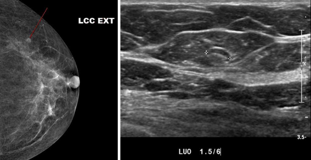
- 6) Intramammary LN : Lentiform mass with central fatty hilum
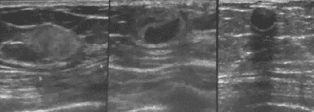
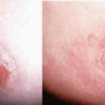
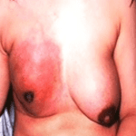
Left) Acute phase : edema of fat
Middle) Subacute phase : complex solid and cystic
Right) Chronic phase : oil cyst, wall calcification