What is the dilated cardiomyopathy (DCMP)
Dilated cardiomyopathy (DCMP) is the most common subtype of nonischemic cardiomyopathy and comprises over 90% of cases.
Dilated cardiomyopathy refers to the final common pathway of a group of diseases rather than a single diagnosis.
In general, there is myocardial dysfunction from progressive dilation and diminishing contractility (systolic dysfunction, FEVG <40%). (Relatively normal diastolic function)
These changes, by definition, are not a result of underlying extramyocardial pathological conditions such as hypertension, coronary artery disease, valvular disease, or congenital abnormalities.
If these conditions are present, given their high prevalence, DCMP should only be diagnosed if the degree of contractile dysfunction is out of proportion to the underlying disease burden.
Histological hallmark of DCMP
Progressive interstitial fibrosis with a numerical decrease of myocyte
Reduced stroke volume ➜ Decreased cardiac output ➜ Congestive heart failure
Main purpose of MRI
– Differentiation from an ischemic origin (DE-MRI)
– Prediction of functional improvement (DE-MRI)
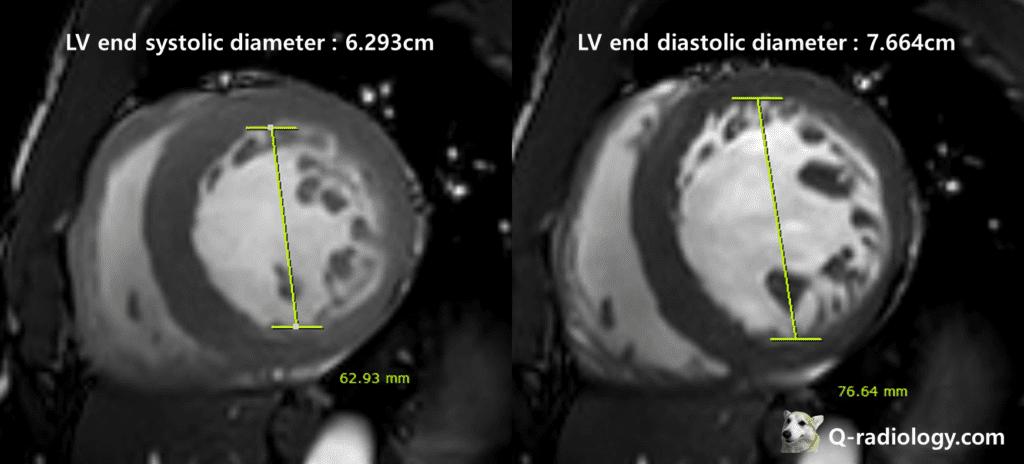
Cine MRI
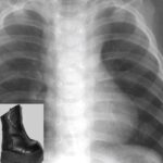
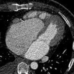
– LV chamber diameter > 6.0cm (at mid-ventricular level)
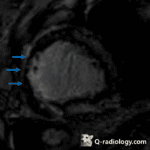
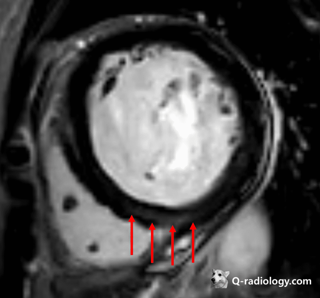
DE-MRI
– Patchy enhancement of longitudinal striae of midwall (28%) or epicardial wall
Worse prognosis than non-enhancement lesions
– Subendocardial, transmural (13%)
– No enhancement (59%)
diffuse thinning of the myocardial wall and dilated left ventricular cavity with associated LV global severe hypokinesia/akinesia
DDx.
– Ischemic heart disease
➜ delayed enhancement at vascular territory
➜ Ischemic heart disease must involve subendocardial wall (subendocardial or transmural)
| Ischemic CMP | Dilated CMP | |
| ECG | Q or ST abnormality | ST or T abnormality |
| Pathology | Necrosis, fibrosis, hibernation | Fibrosis, myocyte size variation |
| Diagnosis | Coronary angiography, Viability study | Coronary angiography, Biopsy |
| DE-MRI | Subendocardial or transmural enhancement | No enhancement Mid wall or patchy enhancement |
Reference)
Charles S. White, Linda B. Haramati, Joseph Jen-Sho Chen, and Jeffrey M. Levsky (2014), Cardiac Imaging, Oxford university press