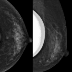
- Amount of fibroglandular tissue
- a) Almost entirely fat
- b) Scattered fibroglandular tissue
- c) Heterogeneous fibroglandular tissue
- d) Extreme fibroglandular tissue

- Background parenchymal enhancement
- Evaluate at 90sec to 3min after contrast injection / Elective MRI should be obtained luteal phase(2 weeks, 7-14days)
- Levels : (A) Minimal, (B) Mild, (C) Moderate, (D) Marked
- Symmetric or Asymmetric

- Focus
- Usually < 5mm
- Nonspecific and too small to be characterized morphologically
- Has no corresponding finding on the postcontrast scan.
- If margin or internal enhancement can be assessed, the finding should be considered a small mass and not a focus
- Favoring malignancy in foucs
- Unique and distinct from the background parenchymal enhancement
- No fatty hilum
- Washout kinetics Significantly increased or new from the prior examination


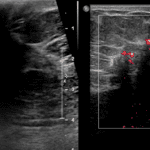
Masses
- Shape / Margin
- Internal enhancement characteristics : homogenous, heterogeneous, rim enhancement, dark internal septations


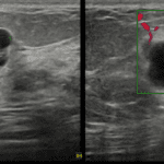
Non-mass enhancement (NME) : Area of enhancement distinct from surrounding parenchyma
- Distribution : Focal, Linear, Segmental, Regional, Multiple regions, Diffuse
- Internal enhancement patterns : Homogenous, Heterogeneous, Clumped, Clustered ring

- Clumped
- Cobblestone, bunch of grapes, string of pearls
- 60% of MR-detected DCIS : Clumped NME
- Clustered ring
- Thin ring around ducts
- Enhancement in periductal stroma
- DCIS
Associated Features
Fat containing lesions
- Lymph nodes, fat necrosis, hamartoma, postoperative seroma/hematoma with fat
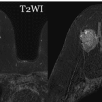
Location of lesion
- Location, Depth

- Initial phase : Change in SI within first two minutes
- Slow <50%
- Medium 50-100%d
- Fast >100%
- Delayed phase
- Persistent
- Plateau
- Washout : decreases<10% after peaking ; 87% are malignant
Implants
- Intracapsular silicone findings (Far more) : Radial folds, Subcapsular line, Keyhole sign (teardrop, noose), Linguine sign
- Linguine sign; multiple curvilinear low-signal-intensity lines floating in silicone gel with no extension beyond the fibrous capsule
- Radial fold -> key hole sign
- Extracapsular silicone : Breast, Lymph nodes
- Rupture of silicone gel-filled breast implant in which the silicone gel is outside of the fibrous scar capsule that forms around the implant.
- Gross high-signal-intensity silicone gel external to the fibrous capsule.