Common in older age than IDC NOS
Slow growing, more favorable prognosis
DCIS are found in 75% of the patients
Imaging finding
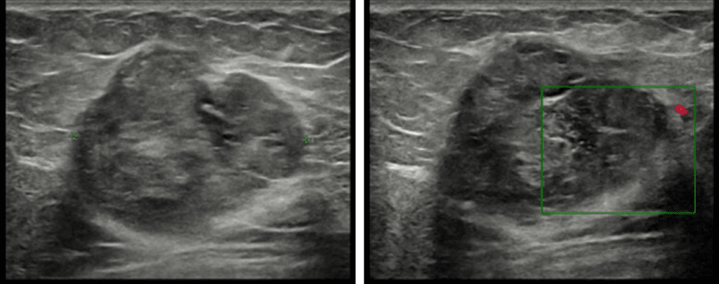
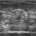
- US : Circumscribed, isoechoic or slightly hypoechoic mass, posterior enhancement > 50%
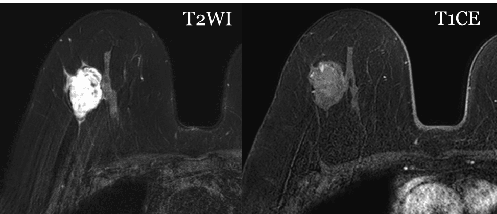
- MR : High signal intensity in T2WI due to mucinous component
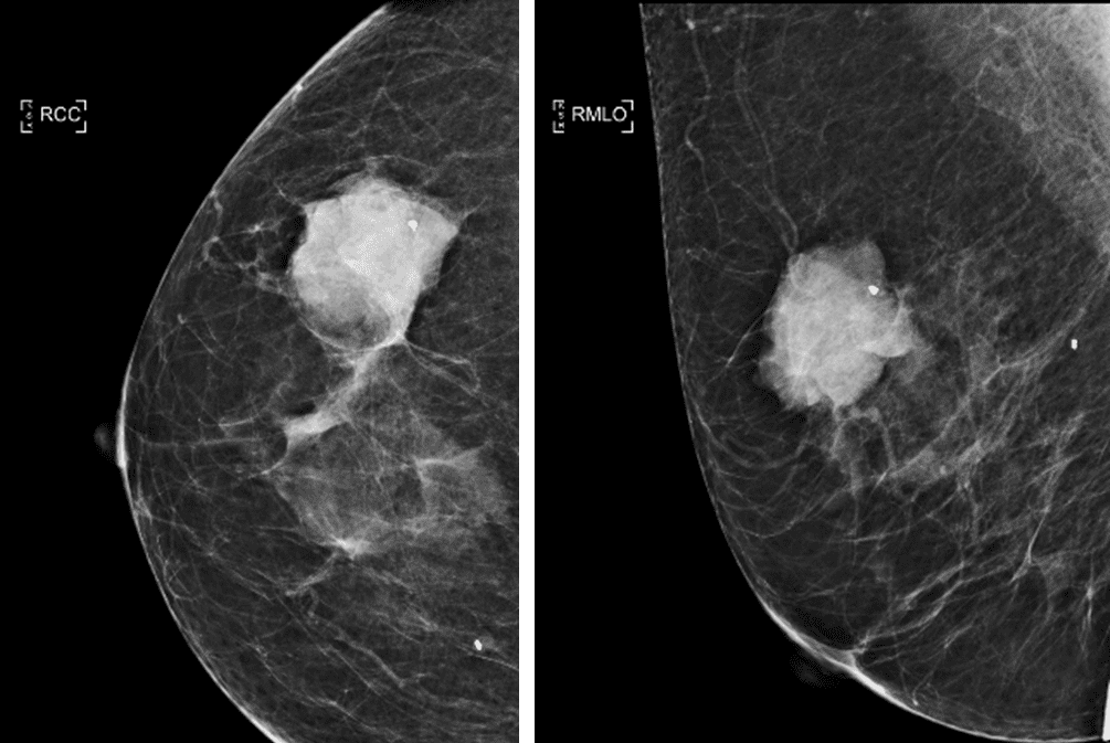
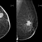
Let’s see a case (83 year-old female) from my hospital which diagnosed as mucinous carcinoma after CNB