Toxoplasmosis in brain(Neruotoxoplasmosis)
Neurotoxoplasmosis is a condition that occurs when the parasite Toxoplasma gondii infects the brain and central nervous system. Toxoplasma gondii is a common parasite that is found in a variety of animals, including cats, birds, and humans. It can be transmitted to humans through contaminated food, water, or soil, or through contact with infected animals.
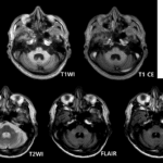
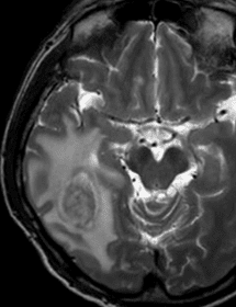
Imaging finding of Toxoplasmosis in brain
- Immunocompromised patient (HIV/AIDS)
- BG, thalamus, corticomedullary junction, cerebellum
- Multifocal > single lesion (15-20%)
- 70% of solitary mass : lymphoma
- Most are small, 2-3cm in diameter
- T2WI : Alternating concentric zones of hyper and hypo SI
- Hyper SI in central necrotizing abscess
- Iso SI in peripheral organizing abscess
- Hyper SI perilesional edema/demyelination
- T1CE : Eccentric target sign
- Reduced rCBC (differential diagnosis with lymphoma)

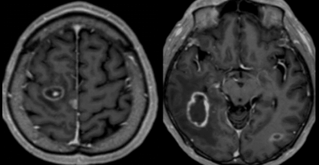
**T1CE : Eccentric target sign**

T2WI : Alternating concentric zones of hyper and hypo SI
- Hyper SI in central necrotizing abscess
- Iso SI in peripheral organizing abscess
- Hyper SI perilesional edema/demyelination
Symptoms of neurotoxoplasmosis
can range from mild to severe and may include fever, fatigue, muscle aches, and headache. In more severe cases, neurotoxoplasmosis can cause seizures, vision loss, and other neurological problems.
Treatment for neurotoxoplasmosis
typically involves the use of medications(sulfadiazine, pyrimethamine) to kill the Toxoplasma gondii parasites. In some cases, steroids may also be used to reduce inflammation in the brain. It is important to seek medical treatment as soon as possible if you suspect you may have neurotoxoplasmosis, as the disease can be difficult to treat if it is not caught in the early stages.
See more about neuroimaging
Follow my instagram