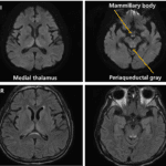
Sturge-Weber syndrome

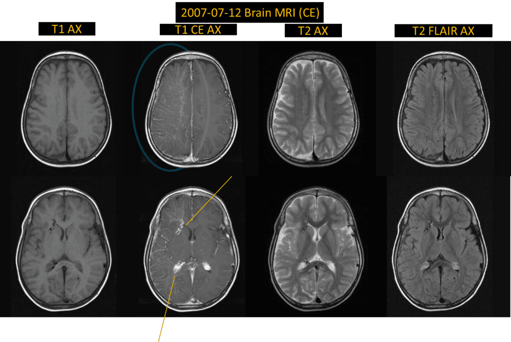
initial MRI showed mild right cerebral atrophy with diffuse leptomeningeal enhancement of right cerebral hemisphere.
medullary vein engorgement and hypertrophied right choroid plexus are found on T2WI and T1CE sequences.
these are typical findings of Sturge-Weber syndrome.

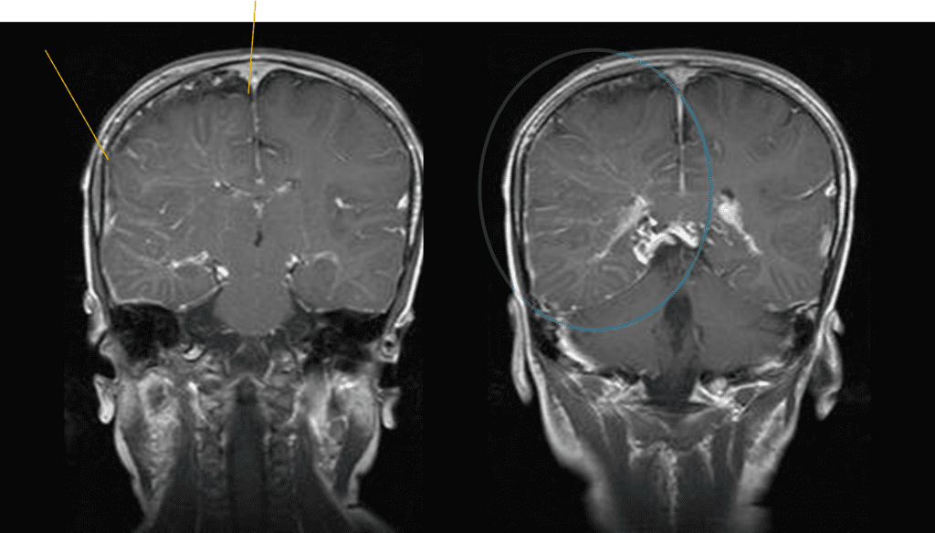


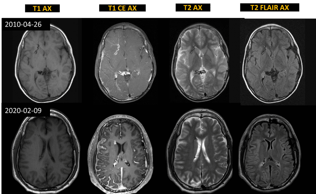
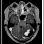
2010) leptomeningeal enhancement along right cerebral cortex with engorgement of medullary vein
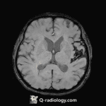
2020) more aggravated atrophic change of right cerebral hemisphere
Sturge-Weber syndrome (=Enecephalotrigeminal angiomatosis)
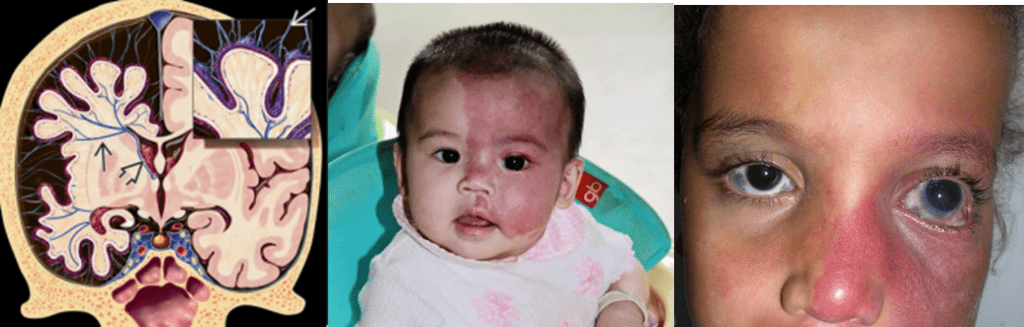
Sturge-weber syndrome is a rare vascular disorder well known for ‘port wine strain’ which is characteristic facial capillary malformation seen at birth.
Reddish-purplish discoloration of skin is found at affected side of face.
Sturge-Weber syndrome is usually sporadic congenital (but not inherited) malformation in which fetal cortical veins fail to develop normally.

Characteristics
- Facial capillary vascular malformation (port wine strain)
- Involving the ophthalmic division (V1) of the trigeminal nerve
- Ipsilateral leptomeningeal angiomatosis
- Angiomatosis of the choroid in ipsilateral eye
- Failure of the primitive venous plexus to regress and mature properly
Clinical manifestation
- Seizure (~1yrs), early onset epilepsy, status epilepticus, development delay
Imaging finding of sturge weber syndrome
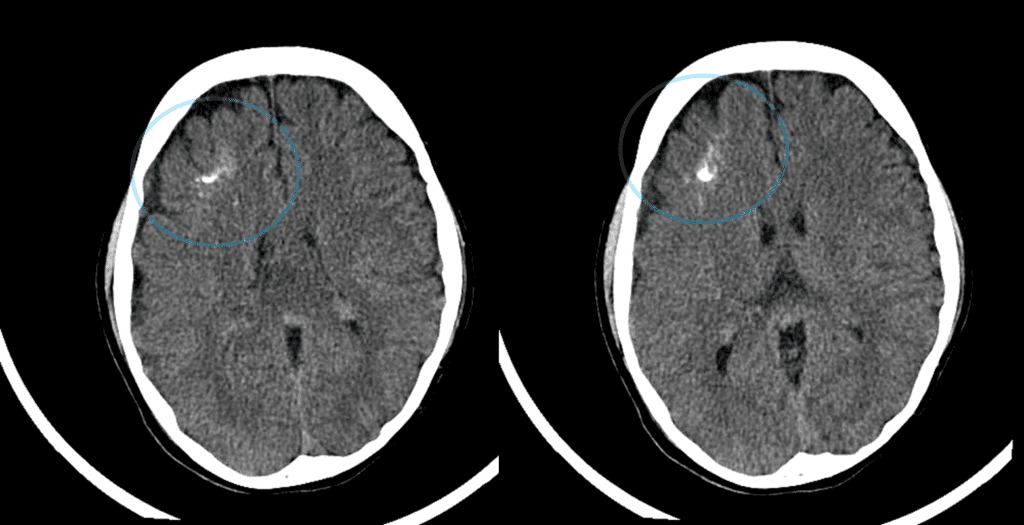
- ‘Tram-track sign‘ on brain CT
- Calcification along cerebral sulcus ➭ reflecting vascular malformation
- Delayed brain atrophy and calcification
- Leptomeningeal (pia mater) vascular malformation (parietal >occipital > frontal lobe)
- Associated with facial capillary malformation
- Abnormal enhancement of pia mater along cerebral sulcus on the same side
➥ “Burnt out” on late phase: decreased pial enhancement
- Ipsilateral choroidal vascular malformation (~70%)
- Compensatory venous drainage
- Especially prominent deep cerebral venous system (medullary vein, subependymal vein …etc)
- Enlarged choroid plexus in the atria

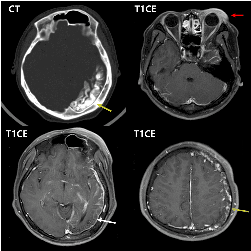
Red arrow – facial capillary malformation
White arrow – left cerebral leptomeningeal enhancement with atrophied left occipital lobe
Green arrow – pial angiomatosis along left cerebral convexities
See more about neuroimaging
Follow my instagram


References)
RadioGraphics 2013; 33:175–195
AJNR Am J Neuroradiol 2009 Feb;30(2):276-81