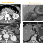
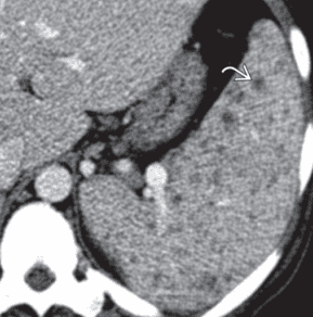
• Pyogenic abscess on CECT
– Low-attenuation complex fluid collection ± air-fluid levels
– Internal gas bubbles, which although uncommon, are very specific for splenic abscess
– Multiloculated appearance seen with liver abscesses possible, but less common with splenic abscess
– May extend to subcapsular location and may rarely cause splenic rupture with generalized peritonitis
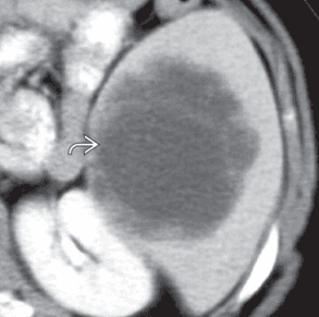
• Fungal microabscesses on CECT
; in setting of immunosuppression, HIV/AIDS, or hematologic disorders
– Multiple small hypodense lesions measuring a few mm
– Multiple punctate splenic calcifications after treatment
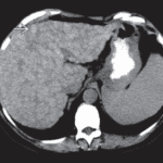
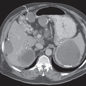
• Echinococcal (hydatid) cyst on CECT
– Complex cyst with multiple low density “daughter” cysts and thick, enhancing wall (“cyst within a cyst”)
– Serpiginous, linear densities within cyst due to collapsed parasitic membranes (water lily sign)
– May demonstrate thick peripheral calcification or internal wavy, curvilinear calcification in chronic setting


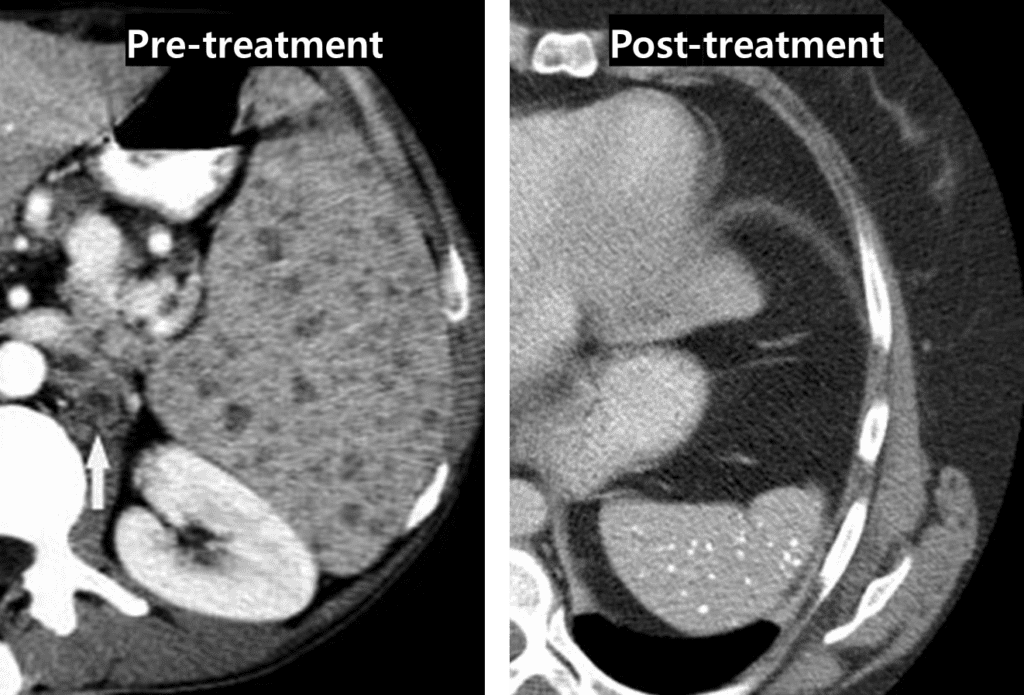
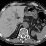
Post-treatment image shows multiple calcifications in spleen
It revealed splenic tuberculosis.
Reference)
Diagnostic Imaging, gastrointestinal 3rd.