Neurocysticercosis
Neurocysticercosis is a disease caused by infection with the pork tapeworm (Taenia solium). It occurs when the larvae of the tapeworm infect the brain, spinal cord, or other tissues in the body. Neurocysticercosis is most commonly found in people who live in or have traveled to areas where the pork tapeworm is prevalent, such as Latin America, Africa, and Asia.


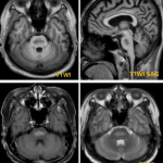
Neurocysticercosis has four pathologic stages : Vesicular, colloidal vesicular, granular nodular, and nodular calcified stage.
- Vesicular stage
- viable cysticercus
- 1~2cm cyst with 1-2mm mural nodule
- mural nodule = scolex of larva
- shows CSF signal on MRI
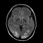
- Colloidal vesicular stage
- Thick cyst wall enhances
- T2 hyperintense cyst with surrounding edema, mild to marked
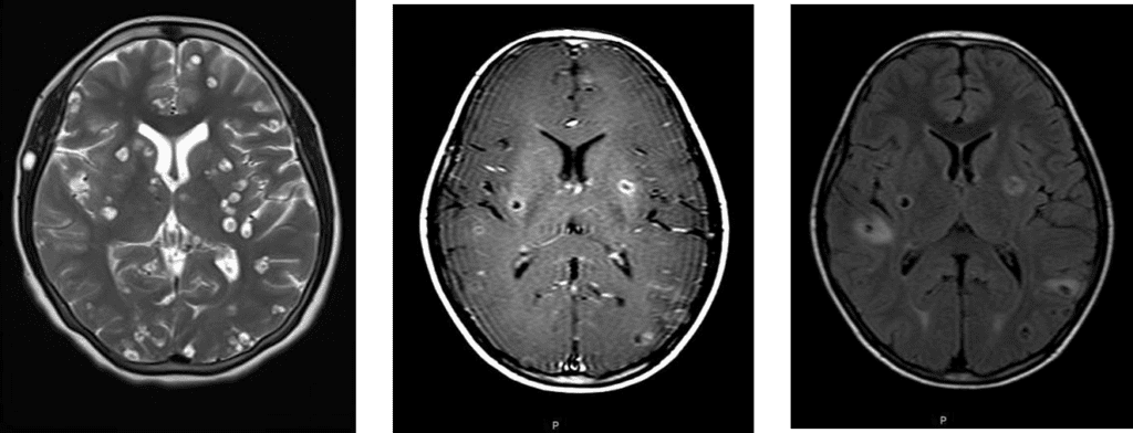
- Granular nodular stage
- Thickened, retracted cyst wall; edema decreases
- About 1cm sized enhancing nodule
- Nodular or ring-like enhancement
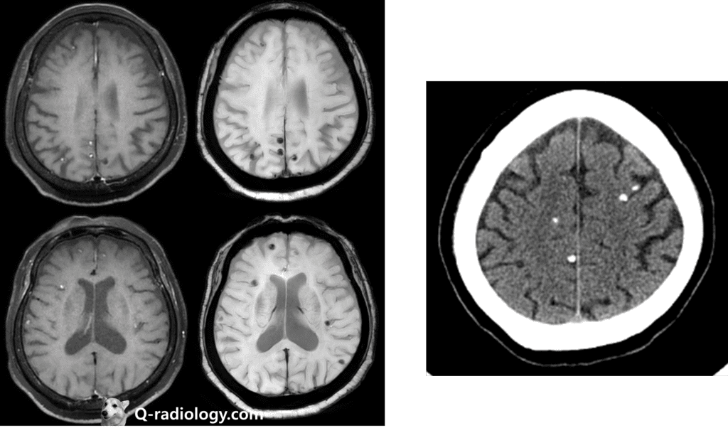
- Nodular calcified stage
- Shrunken, Ca++ lesion
- Rare minimal enhancement


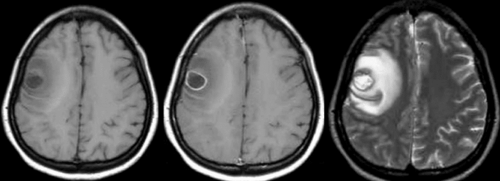
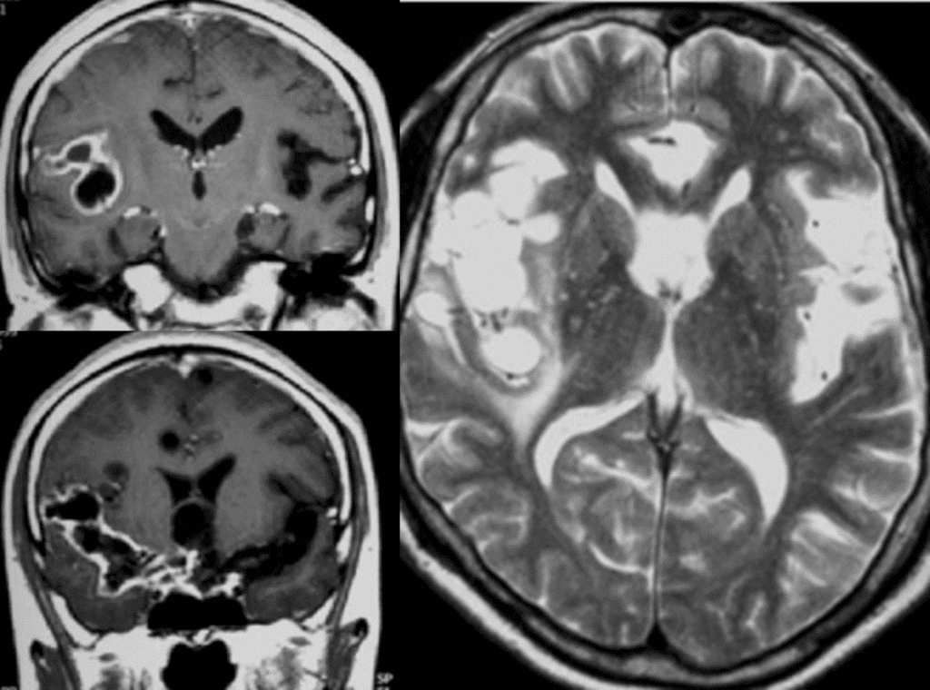
Thick cyst wall enhances with T2 hyperintense cyst with marked surrounding edema, mild to marked

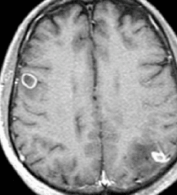
rim enhancing pattern, cyst wall retracted (left occipital lobe)

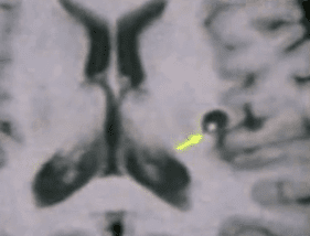
- Cysticercosis : Leptomeningeal form
- Cysticercus located in sulcis and cistern
- Nonvisualization of scolex (no mural nodule)
- Racemose type or large solitary cyst
- Leptomeningeal enhancement – inflammatory reaction at adjacent leptomeninges


Symptoms of neurocysticercosis
can vary widely, depending on the location and number of cysts in the body. Some people may have no symptoms, while others may experience seizures, headaches, visual changes, or other neurological problems. In severe cases, neurocysticercosis can lead to serious complications, including brain damage, hydrocephalus (a build-up of fluid in the brain), and death.
Treatment for neurocysticercosis
typically involves a combination of medications to kill the tapeworm and reduce inflammation in the brain. Surgery may also be necessary to remove cysts or relieve pressure on the brain. It is important to seek medical treatment as soon as possible if you suspect you may have neurocysticercosis, as the disease can be difficult to treat if it is not caught in the early stages.
See more about neuroimaging
Follow my instagram