Etiology of mitral regurgitation
1) Chordae tendinae abnormalities
– congenital mitral valve prolapse (most common)
– chordae tendinae or papillary muscle rupture from trauma, infection, or MI
2) Myxoid degeneration of the leaflets
3) Mitral annular dilatation from LV dilatation
4) Perforation of the leaflets due to infective endocarditis
Pathophysiology
– Depends on compliance of left atrium and pulmonary vascular bed
Acute mitral regurgitation
– Rapid left atrium, left ventricle volume overload
– Acute severe left ventricle failure
– Acute pulmonary edema
Long standing mitral regurgitation
– Left atrium and left ventricle dilatation (adaptation)
– Less severe pulmonary vascular congestion

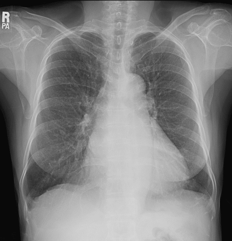
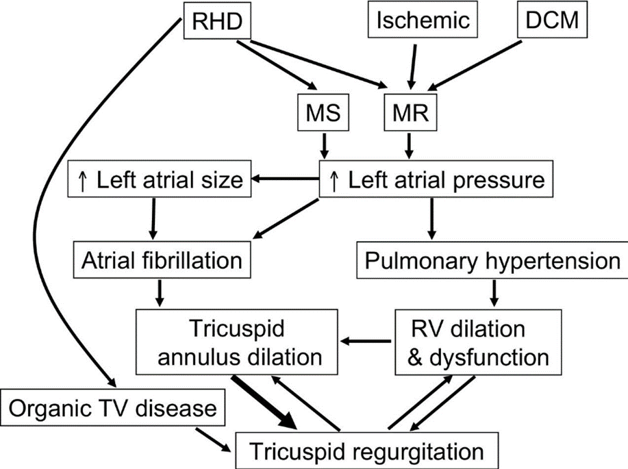
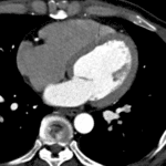
Imaging finding of mitral regurgitation
CT
Thickened leaflets & tendinous chords
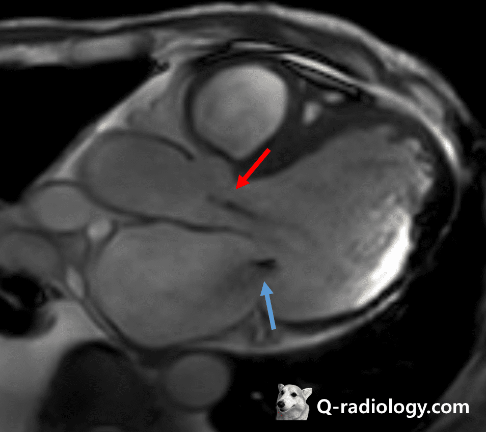
Leaflet prolapse during systolic phase
Mitral annulus calcification
Planimetric regurgitant orifice area
LA and LV enlargement
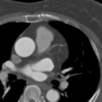
MRI
Signal void in LA
Regurgitant fraction


Tricuspid valve in left-sided valvular disease
– TV annular dilatation and RV enlargement
– Mitral/Aortic valve disease ➜ secondary TR