- Osteonecrosis of lunate bone
- Fixed position in wrist
- Trauma, steroid
- Negative ulnar variance
- 20~40 yr
- Progressive pain, edema, limitation of motion
- Fissure, sclerosis, condensation, fragmentation of lunate bone
- Sequelae (+) : disorganization of wrist, schapholunate dissociation, osteoarthritis
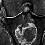
- MR
- Early diagnosis
- TFCC or ligament injury, osteoarthritis

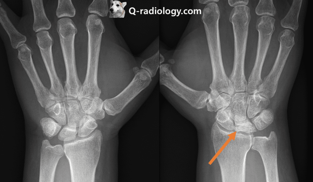
Kienbock’s disease : Fragmentation, collapse, sclerosis of right lunate
Note the negative ulnar variance (the ulna is abnormally shortened, by more than 2.5 mm, compared to the radius)

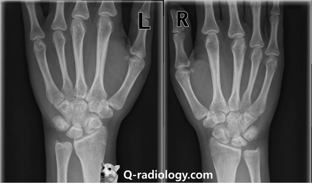
Radiolucency and sclerosis are shown in right lunate with negative ulnar variance, suggestive Kienbock’s disease.

Radiolucency and sclerosis are shown in right lunate (compare with left side lunate) with negative ulnar variance, suggestive Kienbock’s disease.
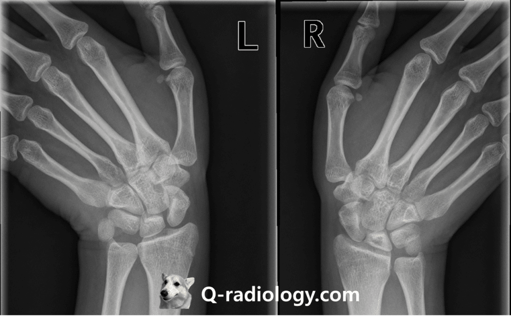
the purpose of this kind of additional view is to free the scaphoid from bony superimposition.

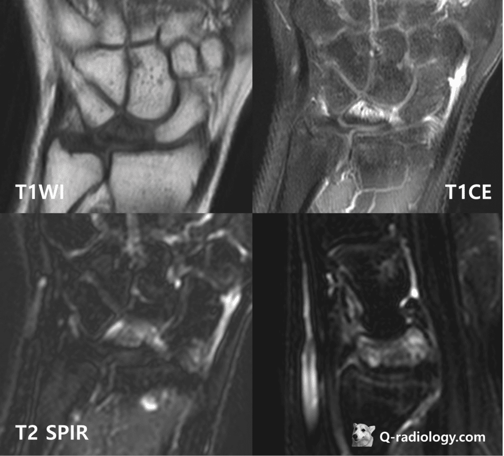
the images reveal edematous change of lunate (low SI on T1WI with enhancement, high signal intensity on T2WI) and sclerosis of lunate (peripheral low signal on T1 and T2WI, but the finding is better visualized on simple radiograph than MRI)