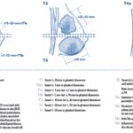
- Infrequent form of invasive breast carcinoma (<1%)
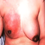
- Increased volume and induration of the breast
- Increased temperature, Tenderness or pain
- Peau d’orange or cutaneous edema, redness of the skin of at least one-third of the breast
- The presence of a palpable ridge at the margin of induration
- Inflammatory breast cancer T stage : T4d
** Primary breast cancer + dilated dermal lymphatic vessels **
Imaging finding
- MG
- diffuse skin thickening, increased density, trabecular thickening, axillary lymphadenopathy with/without associated mass or malignant calcifications
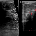
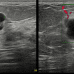
- US
- Marked skin thickening
- Parenchymal acoustic shadowing
- Hypoechogenic areas that did not demonstrate a mass configuration are localized close to the skin
- MR
- Dilatation of subcutaneous lymphatic vessels, edematous change of breast and chest wall
- Optimal for evaluation of neoadjuvant therapy

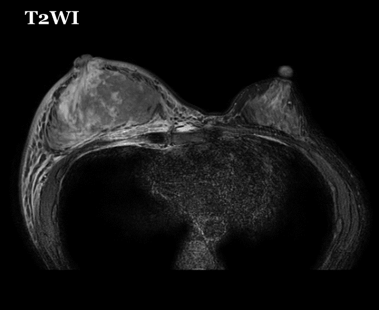
note that markedly increased size of right breast with edematous change in breast and chest wall.
The patient’s mamography was not obtained due to severe pain

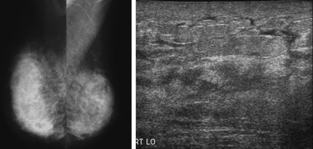
USG : Diffuse skin thickening , increased subcutaneous fat echogenicity and fluid sheet along Cooper’s ligament