Hemangioblastoma
Hemangioblastoma is a type of brain tumor that arises from cells called hemangioblast cells. These cells are found in the blood vessels of the brain and spinal cord and are responsible for the formation of new blood vessels. hemangioblastomas are rare tumors that typically occur in the cerebellum, which is the part of the brain that controls balance and coordination. About 25 to 40% of the tumor occurs in patients with Von Hippel Lindau syndrome.
Imaging finding of Hemangioblastoma
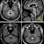
- Benign, slowly growing, vascular neoplasm
- Location
- Posterior fossa (90-95%)
- Cerebellar hemispheres (80%)
- Vermis (15%), medulla or 4th ventricle (5%)
- Supratentorial (5-10%) (around optic pathways, hemispheres; usually in VHL)
- Spinal cord, typically dorsal surface
- Posterior fossa (90-95%)
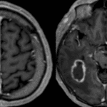
- Morphology
- 50-60% cyst + “mural” nodule ; 40% solid
- Multiple : von Hippel-Lindau syndrome (RCC, pheochromocytoma, retinal tumor, endolymphatic sac tumor)
- CT/MR findings
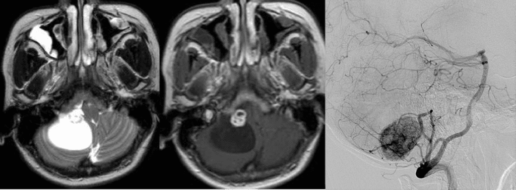
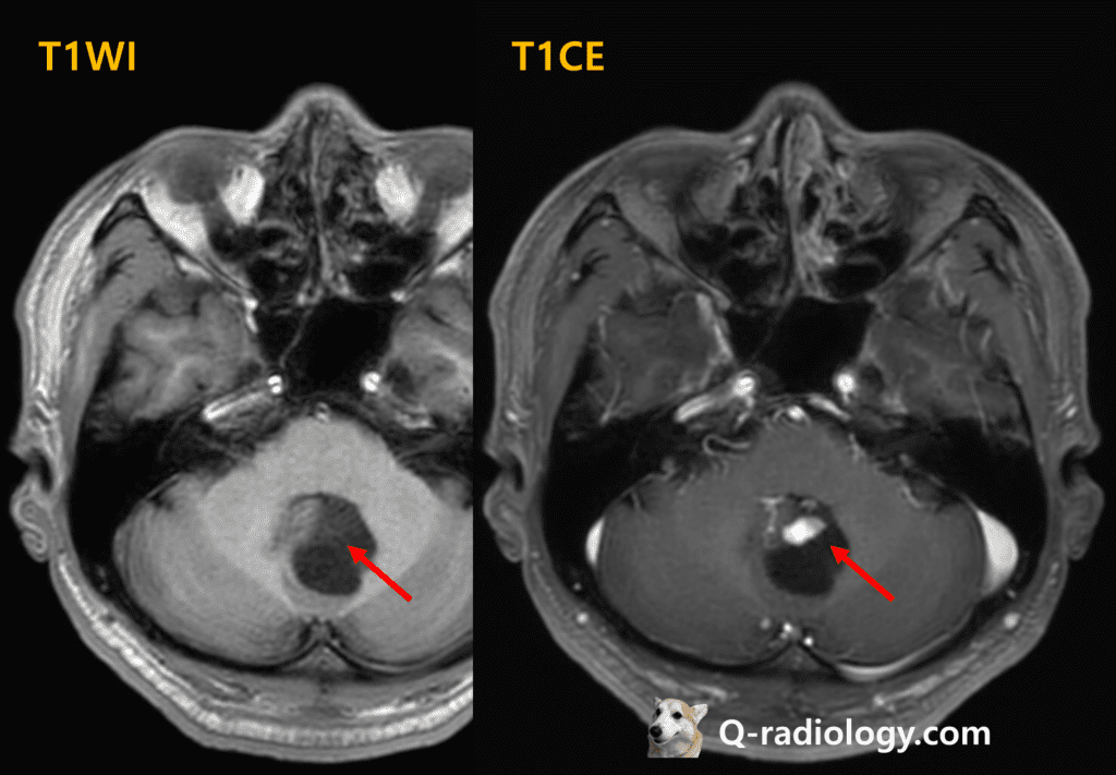
- Cystic mass with strongly enhancing mural nodule
- Angiographic findings
- Large avascular mass (cyst)
- Highly vascular nodule
- Strong and persistent vascular staining of peripheral mural nodule
- Prolonged blush
- with or without arteriovenous shunting (early draining vein)

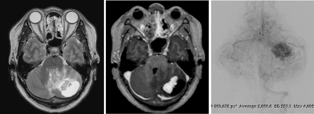
Strong and persistent vascular blush of the tumor on angiography

Strong and persistent vascular blush of the tumor on angiography

See more about neuroimaging
Follow my instagram