Airway obstruction secondary to inflammation of epiglottis & surrounding tissues
IMAGING
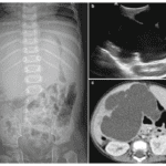
– Lateral radiograph
1. Marked thickening of epiglottis
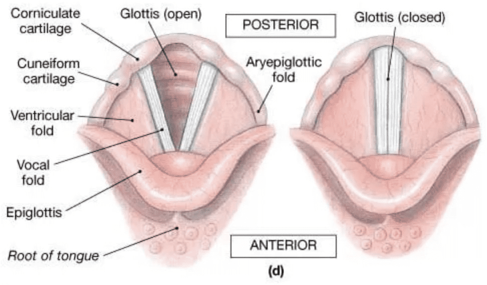
2. Thickening of aryepiglottic folds
– Extend from epiglottis anteriosuperiorly to arytenoid cartilages posteroinferiorly
– Normally thin and convex inferiorly
– May become thickened and convex superiorly
– Swelling of these folds ➜ Actual airway obstruction
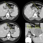
– CT does not play routine role in diagnosing epiglottitis

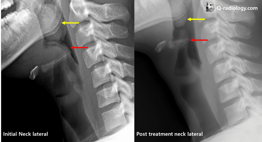
Swollen epiglottis is shown on left image
After treatment the epiglottis’s size is decreased